Zhou Qiankun, Luo Deqing, Li Teng, Liu Zhirong, Zou Weitao, Wang Lei, Lin Dasheng, Lian Kejian
Department of Orthopedic Surgery, The Affiliated Southeast Hospital of Xiamen University, Orthopedic Center of People's Liberation Army, Zhangzhou, Fujian 363000, P.R. China.
Exp Ther Med. 2015 Nov;10(5):1675-1680. doi: 10.3892/etm.2015.2752. Epub 2015 Sep 17.
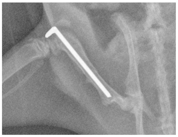
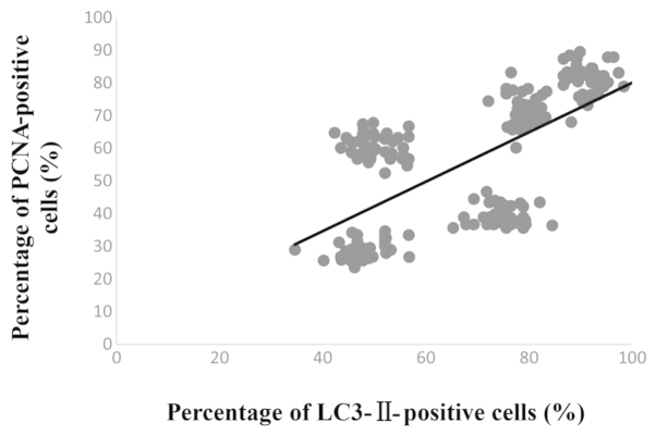
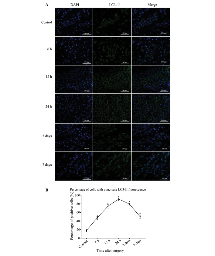
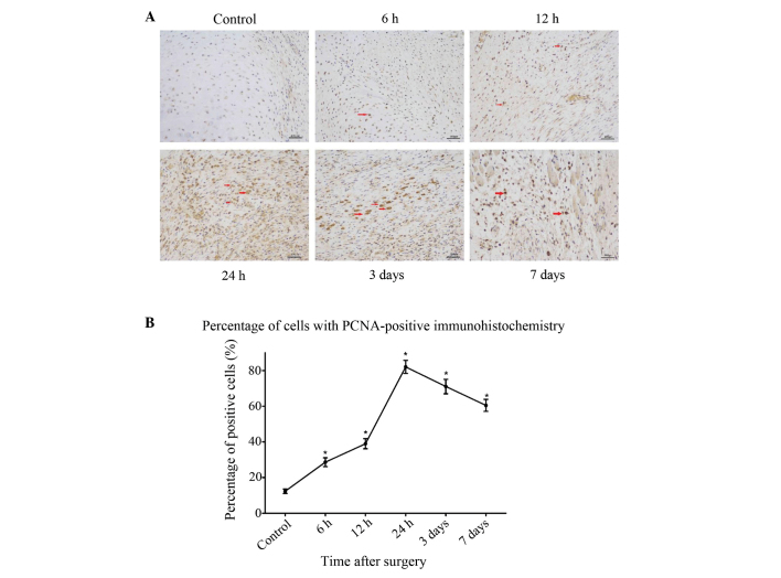
Autophagy, which is a mechanism for the turnover of intracellular molecules and organelles, protects cells during stress responses; however, the role of autophagy in the stages of bone fracture remains to be elucidated. The aim of the present study was to investigate the process of autophagy in bone tissue at different time-points after fracture. A femur fracture model was established in male adult Wistar rats via surgery. The protein expression of microtubule-associated protein II light chain 3 (LC3-II) was analyzed in a femur fracture (experimental) group and a sham-surgery group using immunofluorescence. The protein expression of proliferating cell nuclear antigen (PCNA) was used to investigate the cell proliferation in bone tissue following fracture via immunohistochemical analysis. The correlation between cell proliferation and autophagy was analyzed using linear regression. LC3-II protein was constitutively expressed in the sham-surgery group; however, compared with the expression in the sham-surgery group, the LC3-II expression in the experimental group was significantly increased at each time-point (P<0.05). Similarly, immunohistochemistry revealed that the number of PCNA-positive cells in each section was significantly increased following fracture injury (P<0.01). A comparison of the LC3-II- and PCNA-positive rates in the experimental group rats at each time-point revealed a linear correlation (R=0.43, P<0.01). In conclusion, surgically induced fracture in rats is associated with an increase in LC3-II and PCNA protein expression during the initial stages of fracture injury, and a correlation exists between the expression of the two proteins. These results suggest that potential treatment aimed at improving fracture healing should target the process of autophagy.
自噬是细胞内分子和细胞器更新的一种机制,在应激反应中保护细胞;然而,自噬在骨折各阶段中的作用仍有待阐明。本研究的目的是调查骨折后不同时间点骨组织中的自噬过程。通过手术在成年雄性Wistar大鼠中建立股骨骨折模型。使用免疫荧光法分析股骨骨折(实验组)和假手术组中微管相关蛋白II轻链3(LC3-II)的蛋白表达。通过免疫组织化学分析,使用增殖细胞核抗原(PCNA)的蛋白表达来研究骨折后骨组织中的细胞增殖。使用线性回归分析细胞增殖与自噬之间的相关性。LC3-II蛋白在假手术组中组成性表达;然而,与假手术组中的表达相比,实验组中LC3-II的表达在每个时间点均显著增加(P<0.05)。同样,免疫组织化学显示,骨折损伤后每个切片中PCNA阳性细胞的数量显著增加(P<0.01)。对实验组大鼠在每个时间点的LC3-II和PCNA阳性率进行比较,发现存在线性相关性(R=0.43,P<0.01)。总之,手术诱导的大鼠骨折与骨折损伤初始阶段LC3-II和PCNA蛋白表达增加相关,且这两种蛋白的表达之间存在相关性。这些结果表明,旨在改善骨折愈合的潜在治疗应针对自噬过程。