Williams Rebecca J, Reutens David C, Hocking Julia
Centre for Advanced Imaging The University of Queensland St Lucia Qld 4067 Australia ; Queensland Brain Institute The University of Queensland St Lucia Qld 4067 Australia ; Centre for Clinical Research The University of Queensland Brisbane Qld 4006 Australia ; Hotchkiss Brain Institute and Department of Radiology University of Calgary Calgary AB T2N 4N1 Canada.
Centre for Advanced Imaging The University of Queensland St Lucia Qld 4067 Australia.
Brain Behav. 2015 Oct 14;5(11):e00408. doi: 10.1002/brb3.408. eCollection 2015 Nov.
Decreased water displacement following increased neural activity has been observed using diffusion-weighted functional MRI (DfMRI) at high b-values. The physiological mechanisms underlying the diffusion signal change may be unique from the standard blood oxygenation level-dependent (BOLD) contrast and closer to the source of neural activity. Whether DfMRI reflects neural activity more directly than BOLD outside the primary cerebral regions remains unclear.
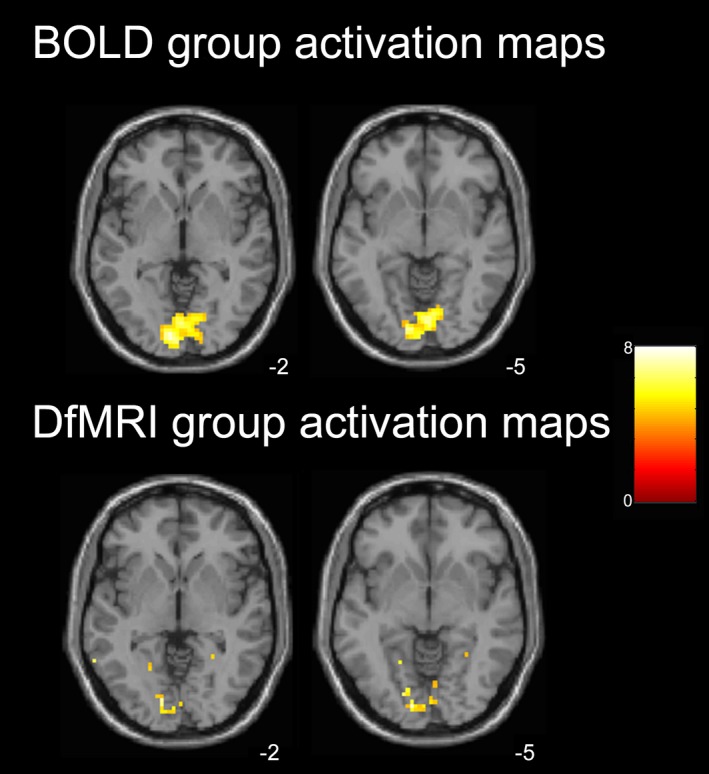
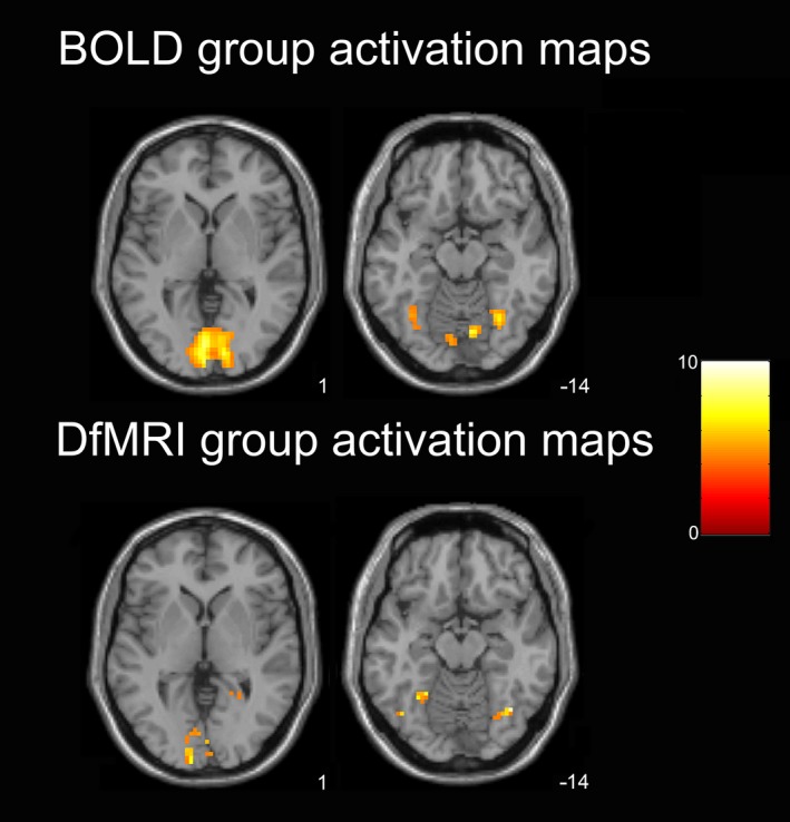
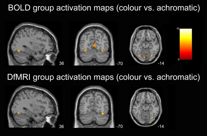
Colored and achromatic Mondrian visual stimuli were statistically contrasted to functionally localize the human color center Area V4 in neurologically intact adults. Spatial and temporal properties of DfMRI and BOLD activation were examined across regions of the visual cortex.
At the individual level, DfMRI activation patterns showed greater spatial specificity to V4 than BOLD. The BOLD activation patterns were more prominent in the primary visual cortex than DfMRI, where activation was localized to the ventral temporal lobe. Temporally, the diffusion signal change in V4 and V1 both preceded the corresponding hemodynamic response, however the early diffusion signal change was more evident in V1.
DfMRI may be of use in imaging applications implementing cognitive subtraction paradigms, and where highly precise individual functional localization is required.
使用高b值的扩散加权功能磁共振成像(DfMRI)观察到,神经活动增加后水位移减少。扩散信号变化背后的生理机制可能与标准的血氧水平依赖(BOLD)对比不同,且更接近神经活动的源头。在初级脑区之外,DfMRI是否比BOLD更直接地反映神经活动仍不清楚。
对彩色和无色蒙德里安视觉刺激进行统计学对比,以在神经功能完好的成年人中对人类颜色中心V4区进行功能定位。在视觉皮层各区域检查DfMRI和BOLD激活的空间和时间特性。
在个体水平上,DfMRI激活模式对V4区的空间特异性高于BOLD。BOLD激活模式在初级视觉皮层比DfMRI更显著,DfMRI激活定位于颞叶腹侧。在时间上,V4区和V1区的扩散信号变化均先于相应的血液动力学反应,然而早期扩散信号变化在V1区更明显。
DfMRI可能在实施认知减法范式的成像应用以及需要高精度个体功能定位的情况下有用。