Berman Sara E, Rivera-Rivera Leonardo A, Clark Lindsay R, Racine Annie M, Keevil Jon G, Bratzke Lisa C, Carlsson Cynthia M, Bendlin Barbara B, Rowley Howard A, Blennow Kaj, Zetterberg Henrik, Asthana Sanjay, Turski Patrick, Johnson Sterling C, Wieben Oliver
Alzheimer's Disease Research Center, University of Wisconsin School of Medicine and Public Health, Madison, WI 53792 ; Neuroscience Training Program, University of Wisconsin-Madison, Madison, WI 53705 ; Medical Scientist Training Program, University of Wisconsin-Madison, Madison, WI 53705.
Department of Medical Physics, University of Wisconsin School of Medicine and Public Health, Madison, WI 53705.
Alzheimers Dement (Amst). 2015 Dec 1;1(4):420-428. doi: 10.1016/j.dadm.2015.09.005.
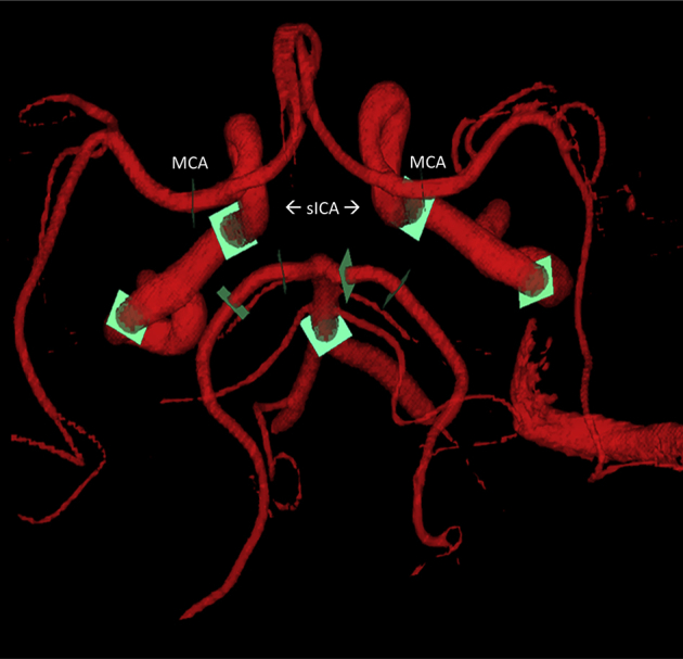
While cerebrovascular disease has long been known to co-occur with Alzheimer's disease (AD), recent studies suggest an etiologic contribution to AD pathogenesis. We used 4D-Flow magnetic resonance imaging (MRI) to evaluate blood flow and pulsatility indices in the Circle of Willis. We hypothesized decreased mean blood flow and increased pulsatility, metrics indicative of poor vascular health, would be associated with cerebral atrophy and an AD cerebrospinal fluid (CSF) profile.
312 patients along the AD continuum (172 middle-aged, 60 cognitively-healthy older, 44 mild cognitive impairment (MCI), and 36 AD) underwent MRI, CSF, and medical examinations. Regression was used to predict CSF biomarkers and atrophy from 4D-Flow, and ANCOVA to compare vascular health between groups.
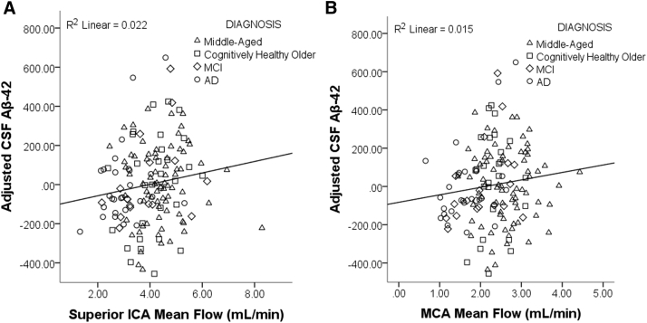
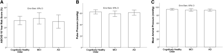
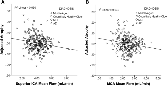
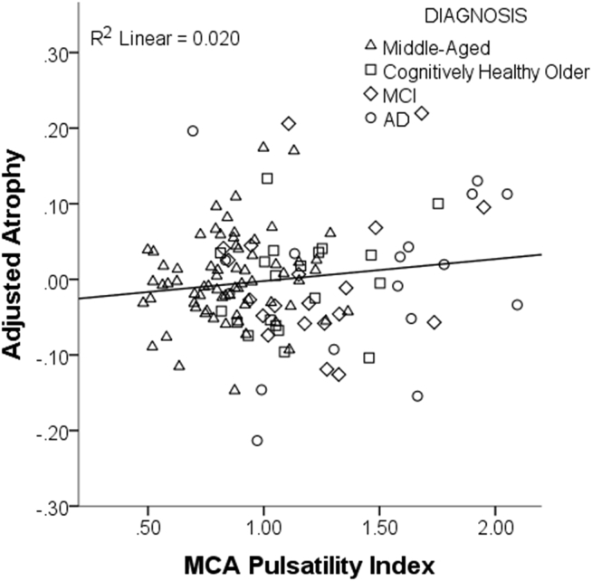
Decreased mean flow in the middle cerebral (MCA) and superior portion of the internal carotid arteries (sICA) and increased pulsatility in the MCA were associated with greater brain atrophy. Decreased mean flow in the sICA was associated with lower Aβ-42 in the CSF, a pathological biomarker profile associated with AD. Interestingly, although metrics of flow and pulsatility differed markedly across the AD spectrum, there were no significant differences in cardiovascular risk score, mean arterial pressure and pulse pressure across the three age-matched older cohorts.
By measuring intracranial arterial health directly with 4D-Flow MRI, these data suggest that intracranial arterial health is compromised in symptomatic AD. Even after accounting for disease stage, cerebral artery health is associated with atrophy and an AD Aβ-42 profile, suggesting neurovascular health may contribute to the etiopathogenesis of AD.
虽然长期以来人们都知道脑血管疾病与阿尔茨海默病(AD)共同出现,但最近的研究表明其对AD发病机制有病因学贡献。我们使用四维血流磁共振成像(MRI)来评估 Willis 环中的血流和搏动指数。我们假设平均血流减少和搏动增加(这些指标表明血管健康状况不佳)会与脑萎缩和AD脑脊液(CSF)特征相关。
312名处于AD连续体中的患者(172名中年人、60名认知健康的老年人、44名轻度认知障碍(MCI)患者和36名AD患者)接受了MRI、CSF和医学检查。使用回归分析从四维血流预测CSF生物标志物和萎缩情况,并使用协方差分析比较各组之间的血管健康状况。
大脑中动脉(MCA)和颈内动脉上段(sICA)的平均血流减少以及MCA搏动增加与更严重的脑萎缩相关。sICA平均血流减少与CSF中较低的Aβ - 42相关,Aβ - 42是与AD相关的病理生物标志物特征。有趣的是,尽管在AD谱系中血流和搏动指标有显著差异,但在三个年龄匹配的老年队列中,心血管风险评分、平均动脉压和脉压没有显著差异。
通过使用四维血流MRI直接测量颅内动脉健康状况,这些数据表明有症状的AD患者颅内动脉健康受损。即使考虑疾病阶段,脑动脉健康仍与萎缩和AD的Aβ - 42特征相关,这表明神经血管健康可能对AD的病因发病机制有影响。