• 文献检索
  • 文档翻译
  • 深度研究
  • 学术资讯
  • Suppr Zotero 插件Zotero 插件
  • 邀请有礼
  • 套餐&价格
  • 历史记录
应用&插件
Suppr Zotero 插件Zotero 插件浏览器插件Mac 客户端Windows 客户端微信小程序
定价
高级版会员购买积分包购买API积分包
服务
文献检索文档翻译深度研究API 文档MCP 服务
关于我们
关于 Suppr公司介绍联系我们用户协议隐私条款
关注我们

Suppr 超能文献

核心技术专利:CN118964589B侵权必究
粤ICP备2023148730 号-1Suppr @ 2026

文献检索

告别复杂PubMed语法,用中文像聊天一样搜索,搜遍4000万医学文献。AI智能推荐,让科研检索更轻松。

立即免费搜索

文件翻译

保留排版,准确专业,支持PDF/Word/PPT等文件格式,支持 12+语言互译。

免费翻译文档

深度研究

AI帮你快速写综述,25分钟生成高质量综述,智能提取关键信息,辅助科研写作。

立即免费体验

方法学及体表面积校正对主动脉根部和弓部超声心动图评估的影响:英国超声心动图学会研究与审计小组委员会的一项研究

Impact of methodology and the use of allometric scaling on the echocardiographic assessment of the aortic root and arch: a study by the Research and Audit Sub-Committee of the British Society of Echocardiography.

作者信息

Oxborough David, Ghani Saqib, Harkness Allan, Lloyd Guy, Moody William, Ring Liam, Sandoval Julie, Senior Roxy, Sheikh Nabeel, Stout Martin, Utomi Victor, Willis James, Zaidi Abbas, Steeds Richard

机构信息

Research Institute for Sports and Exercise Sciences, Liverpool John Moores University , Liverpool, L3 9UT , UK.

Department of Cardiology, Southend University Hospital NHS Trust , Westcliffe on Sea , UK.

出版信息

Echo Res Pract. 2014 Sep 1;1(1):1-9. doi: 10.1530/ERP-14-0004. Epub 2014 May 6.

DOI:10.1530/ERP-14-0004
PMID:26693286
原文链接:https://pmc.ncbi.nlm.nih.gov/articles/PMC4676464/
Abstract

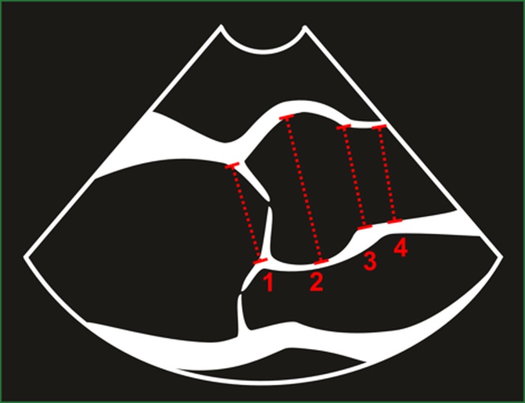
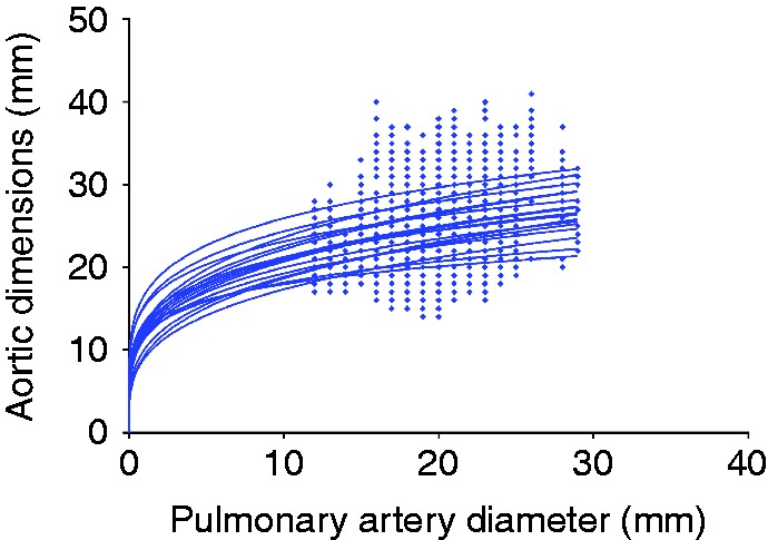
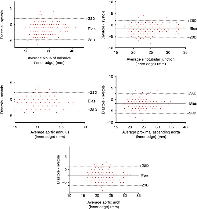
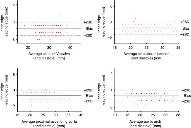
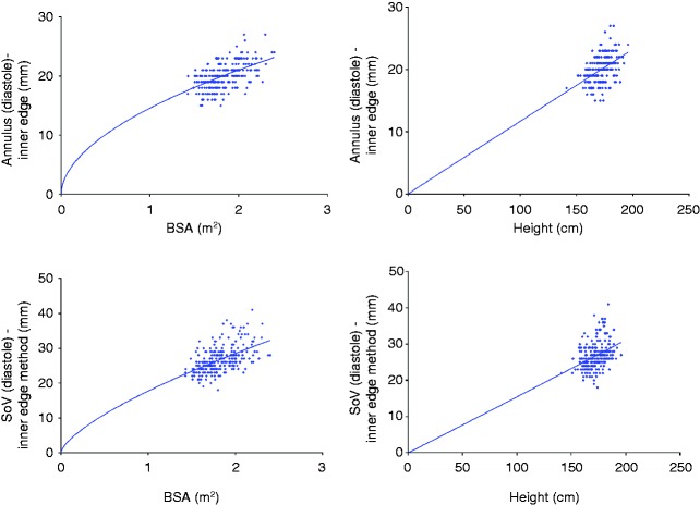
The aim of the study is to establish the impact of 2D echocardiographic methods on absolute values for aortic root dimensions and to describe any allometric relationship to body size. We adopted a nationwide cross-sectional prospective multicentre design using images obtained from studies utilising control groups or where specific normality was being assessed. A total of 248 participants were enrolled with no history of cardiovascular disease, diabetes, hypertension or abnormal findings on echocardiography. Aortic root dimensions were measured at the annulus, the sinus of Valsalva, the sinotubular junction, the proximal ascending aorta and the aortic arch using the inner edge and leading edge methods in both diastole and systole by 2D echocardiography. All dimensions were scaled allometrically to body surface area (BSA), height and pulmonary artery diameter. For all parameters with the exception of the aortic annulus, dimensions were significantly larger in systole (P<0.05). All aortic root and arch measurements were significantly larger when measured using the leading edge method compared with the inner edge method (P<0.05). Allometric scaling provided a b exponent of BSA(0.6) in order to achieve size independence. Similarly, ratio scaling to height in subjects under the age of 40 years also produced size independence. In conclusion, the largest aortic dimensions occur in systole while using the leading edge method. Reproducibility of measurement, however, is better when assessing aortic dimensions in diastole. There is an allometric relationship to BSA and, therefore, allometric scaling in the order of BSA(0.6) provides a size-independent index that is not influenced by the age or gender.

摘要

本研究的目的是确定二维超声心动图方法对主动脉根部尺寸绝对值的影响,并描述其与身体大小的任何异速生长关系。我们采用了全国性横断面前瞻性多中心设计,使用从有对照组的研究或评估特定正常情况的研究中获取的图像。共有248名无心血管疾病、糖尿病、高血压病史且超声心动图检查无异常发现的参与者入组。通过二维超声心动图,在舒张期和收缩期使用内缘法和前缘法测量主动脉根部在瓣环、主动脉窦、窦管交界处、升主动脉近端和主动脉弓处的尺寸。所有尺寸均根据身体表面积(BSA)、身高和肺动脉直径进行异速生长缩放。除主动脉瓣环外,所有参数在收缩期的尺寸均显著更大(P<0.05)。与内缘法相比,使用前缘法测量时,所有主动脉根部和弓部的测量值均显著更大(P<0.05)。为了实现尺寸独立性,异速生长缩放得出BSA的b指数为0.6。同样,40岁以下受试者按身高进行比例缩放也能实现尺寸独立性。总之,使用前缘法时,主动脉最大尺寸出现在收缩期。然而,在舒张期评估主动脉尺寸时测量的可重复性更好。与BSA存在异速生长关系,因此,按BSA(0.6)顺序进行异速生长缩放可提供一个不受年龄或性别影响的尺寸独立指数。

https://cdn.ncbi.nlm.nih.gov/pmc/blobs/3943/4676464/653c7388262a/echo-01-1-g006.jpg
https://cdn.ncbi.nlm.nih.gov/pmc/blobs/3943/4676464/e645cffa3934/echo-01-1-g001.jpg
https://cdn.ncbi.nlm.nih.gov/pmc/blobs/3943/4676464/4e699bd4ca2a/echo-01-1-g002.jpg
https://cdn.ncbi.nlm.nih.gov/pmc/blobs/3943/4676464/ad7c80ceaa5b/echo-01-1-g003.jpg
https://cdn.ncbi.nlm.nih.gov/pmc/blobs/3943/4676464/bae803ba3aed/echo-01-1-g004.jpg
https://cdn.ncbi.nlm.nih.gov/pmc/blobs/3943/4676464/4b057507e0fd/echo-01-1-g005.jpg
https://cdn.ncbi.nlm.nih.gov/pmc/blobs/3943/4676464/653c7388262a/echo-01-1-g006.jpg

相似文献

1
Impact of methodology and the use of allometric scaling on the echocardiographic assessment of the aortic root and arch: a study by the Research and Audit Sub-Committee of the British Society of Echocardiography.方法学及体表面积校正对主动脉根部和弓部超声心动图评估的影响:英国超声心动图学会研究与审计小组委员会的一项研究
Echo Res Pract. 2014 Sep 1;1(1):1-9. doi: 10.1530/ERP-14-0004. Epub 2014 May 6.
2
Scaling to produce size-independent indices of echocardiographic derived aortic root dimensions in elite Rugby Football League players.对精英橄榄球联盟球员进行缩放处理,以生成与尺寸无关的超声心动图衍生主动脉根部尺寸指数。
Ultrasound. 2019 May;27(2):94-100. doi: 10.1177/1742271X18818607. Epub 2019 Jan 17.
3
Two-dimensional transthoracic echocardiographic normal reference ranges for proximal aorta dimensions: results from the EACVI NORRE study.二维经胸超声心动图测定近端主动脉内径的正常参考范围:EACVI NORRE研究结果
Eur Heart J Cardiovasc Imaging. 2017 Feb;18(2):167-179. doi: 10.1093/ehjci/jew053. Epub 2016 Apr 3.
4
Ascending aorta diameters measured by echocardiography using both leading edge-to-leading edge and inner edge-to-inner edge conventions in healthy volunteers.超声心动图测量健康志愿者升主动脉直径的前缘-前缘和内缘-内缘两种方法。
Eur Heart J Cardiovasc Imaging. 2014 Apr;15(4):415-22. doi: 10.1093/ehjci/jet173. Epub 2013 Oct 4.
5
Effects of aging and body size on proximal and ascending aorta and aortic arch: inner edge-to-inner edge reference values in a large adult population by two-dimensional transthoracic echocardiography.二维经胸超声心动图在大型成人人群中对近端和升主动脉及主动脉弓的年龄和体型影响:内缘到内缘参考值。
J Am Soc Echocardiogr. 2013 Apr;26(4):419-27. doi: 10.1016/j.echo.2012.12.013. Epub 2013 Jan 18.
6
Aortic root remodelling in competitive athletes.竞技运动员的主动脉根部重构。
Eur J Prev Cardiol. 2020 Sep;27(14):1518-1526. doi: 10.1177/2047487319894882. Epub 2019 Dec 18.
7
Comparative measurement of aortic root by transthoracic echocardiography in normal Korean population based on two different guidelines.基于两种不同指南,经胸超声心动图对正常韩国人群主动脉根部的比较测量。
Cardiovasc Ultrasound. 2013 Aug 13;11:28. doi: 10.1186/1476-7120-11-28.
8
Normal Values and Differences in Ascending Aortic Diameter in a Healthy Population of Adults as Measured by the Pediatric versus Adult American Society of Echocardiography Guidelines.根据美国超声心动图学会儿科与成人指南测量的健康成年人群升主动脉直径的正常数值及差异
J Am Soc Echocardiogr. 2016 Feb;29(2):166-72. doi: 10.1016/j.echo.2015.09.010. Epub 2015 Oct 23.
9
Transthoracic echocardiographic reference values of the aortic root: results from the Hamburg City Health Study.经胸超声心动图主动脉根部参考值:汉堡城市健康研究结果。
Int J Cardiovasc Imaging. 2021 Dec;37(12):3513-3524. doi: 10.1007/s10554-021-02354-5. Epub 2021 Jul 29.
10
Normal Values of Aortic Root Size According to Age, Sex, and Race: Results of the World Alliance of Societies of Echocardiography Study.根据年龄、性别和种族的主动脉根部大小正常值:世界超声心动图学会联盟研究的结果。
J Am Soc Echocardiogr. 2022 Mar;35(3):267-274. doi: 10.1016/j.echo.2021.09.011. Epub 2021 Oct 4.

引用本文的文献

1
Adverse Thoracic Aortic Remodeling in Obstructive Sleep Apnea.阻塞性睡眠呼吸暂停患者的胸主动脉不良重塑
Am J Hypertens. 2022 Jun 16;35(6):510-511. doi: 10.1093/ajh/hpac019.
2
Normal Values of Aortic Root Size According to Age, Sex, and Race: Results of the World Alliance of Societies of Echocardiography Study.根据年龄、性别和种族的主动脉根部大小正常值:世界超声心动图学会联盟研究的结果。
J Am Soc Echocardiogr. 2022 Mar;35(3):267-274. doi: 10.1016/j.echo.2021.09.011. Epub 2021 Oct 4.
3
Image-based scaling laws for somatic growth and pulmonary artery morphometry from infancy to adulthood.

本文引用的文献

1
Comparative measurement of aortic root by transthoracic echocardiography in normal Korean population based on two different guidelines.基于两种不同指南,经胸超声心动图对正常韩国人群主动脉根部的比较测量。
Cardiovasc Ultrasound. 2013 Aug 13;11:28. doi: 10.1186/1476-7120-11-28.
2
Effects of aging and body size on proximal and ascending aorta and aortic arch: inner edge-to-inner edge reference values in a large adult population by two-dimensional transthoracic echocardiography.二维经胸超声心动图在大型成人人群中对近端和升主动脉及主动脉弓的年龄和体型影响:内缘到内缘参考值。
J Am Soc Echocardiogr. 2013 Apr;26(4):419-27. doi: 10.1016/j.echo.2012.12.013. Epub 2013 Jan 18.
3
基于图像的体部生长和肺动脉形态计量学从婴儿期到成年期的比例定律。
Am J Physiol Heart Circ Physiol. 2020 Aug 1;319(2):H432-H442. doi: 10.1152/ajpheart.00123.2020. Epub 2020 Jul 3.
4
Normal reference intervals for cardiac dimensions and function for use in echocardiographic practice: a guideline from the British Society of Echocardiography.超声心动图检查中使用的心脏大小和功能的正常参考区间:英国超声心动图学会指南
Echo Res Pract. 2020 Feb 24;7(1):G1-G18. doi: 10.1530/ERP-19-0050.
5
Scaling to produce size-independent indices of echocardiographic derived aortic root dimensions in elite Rugby Football League players.对精英橄榄球联盟球员进行缩放处理,以生成与尺寸无关的超声心动图衍生主动脉根部尺寸指数。
Ultrasound. 2019 May;27(2):94-100. doi: 10.1177/1742271X18818607. Epub 2019 Jan 17.
6
Establishment of reference values of the caudal vena cava by fast-ultrasonography through different views in healthy dogs.通过快速超声检查在健康犬不同视图下建立尾静脉腔的参考值。
J Vet Intern Med. 2018 Jul;32(4):1308-1318. doi: 10.1111/jvim.15136. Epub 2018 May 10.
7
Our obsession with normal values.我们对正常数值的痴迷。
Echo Res Pract. 2018 Jun 1;5(2):R17-R21. doi: 10.1530/ERP-17-0082.
8
Right to Left Ventricular Diameter Ratio ≥0.42 is the Warning Flag for Suspecting Atrial Septal Defect in Preschool Children: Age- and Body Surface Area-Related Reference Values Determined by M-Mode Echocardiography.右心室与左心室直径比值≥0.42是学龄前儿童疑似房间隔缺损的警示标志:通过M型超声心动图确定的与年龄和体表面积相关的参考值
Pediatr Cardiol. 2016 Apr;37(4):704-13. doi: 10.1007/s00246-015-1334-y. Epub 2015 Dec 24.
Normal limits in relation to age, body size and gender of two-dimensional echocardiographic aortic root dimensions in persons ≥15 years of age.
二维超声心动图主动脉根部在年龄、体型和性别方面的正常参考值范围:年龄≥15 岁人群。
Am J Cardiol. 2012 Oct 15;110(8):1189-94. doi: 10.1016/j.amjcard.2012.05.063. Epub 2012 Jul 6.
4
Multimodality imaging of aortic dimensions: comparison of transthoracic echocardiography with multidetector row computed tomography.主动脉尺寸的多模态成像:经胸超声心动图与多排螺旋计算机断层扫描的比较
Echocardiography. 2012 Jul;29(6):735-41. doi: 10.1111/j.1540-8175.2012.01666.x. Epub 2012 Mar 9.
5
Assessment of aortic root dimensions in patients with suspected Marfan syndrome: intraindividual comparison of contrast-enhanced and non-contrast magnetic resonance angiography with echocardiography.评估疑似马凡综合征患者的主动脉根部尺寸:对比增强和非对比磁共振血管造影与超声心动图的个体内比较。
Int J Cardiol. 2013 Jul 15;167(1):190-6. doi: 10.1016/j.ijcard.2011.12.041. Epub 2012 Jan 10.
6
The right ventricle of the endurance athlete: the relationship between morphology and deformation.耐力运动员的右心室:形态与变形之间的关系。
J Am Soc Echocardiogr. 2012 Mar;25(3):263-71. doi: 10.1016/j.echo.2011.11.017. Epub 2011 Dec 14.
7
Nomograms for aortic root diameters in children using two-dimensional echocardiography.二维超声心动图测量儿童主动脉根部直径的列线图
Am J Cardiol. 2010 Mar 15;105(6):888-94. doi: 10.1016/j.amjcard.2009.11.040.
8
CT and MRI in diseases of the aorta.主动脉疾病的CT与MRI检查
AJR Am J Roentgenol. 2009 Oct;193(4):928-40. doi: 10.2214/AJR.08.2166.
9
Assessment of the thoracic aorta by multidetector computed tomography: age- and sex-specific reference values in adults without evident cardiovascular disease.多排螺旋计算机断层扫描评估胸主动脉:无明显心血管疾病成年人的年龄和性别特异性参考值
J Cardiovasc Comput Tomogr. 2008 Sep-Oct;2(5):298-308. doi: 10.1016/j.jcct.2008.08.002. Epub 2008 Aug 7.
10
Longitudinal plane colour tissue-Doppler myocardial velocities and their association with left ventricular length, volume, and mass in humans.纵向平面彩色组织多普勒心肌速度及其与人类左心室长度、容积和质量的关系。
Eur J Echocardiogr. 2008 Jul;9(4):542-6. doi: 10.1093/ejechocard/jen114. Epub 2008 Mar 11.