Joshi Bishnu P, Zhou Juan, Pant Asha, Duan Xiyu, Zhou Quan, Kuick Rork, Owens Scott R, Appelman Henry, Wang Thomas D
Department of Medicine, Division of Gastroenterology, ‡Department of Biomedical Engineering, §Department of Biostatistics, ∥Department of Pathology, and ⊥Department of Mechanical Engineering, University of Michigan , Ann Arbor, Michigan 48109, United States.
Bioconjug Chem. 2016 Feb 17;27(2):481-94. doi: 10.1021/acs.bioconjchem.5b00565. Epub 2015 Dec 28.
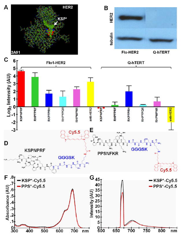
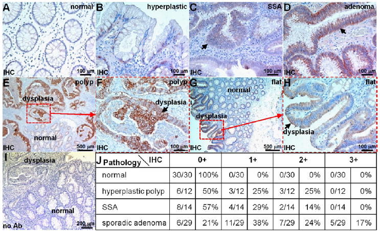
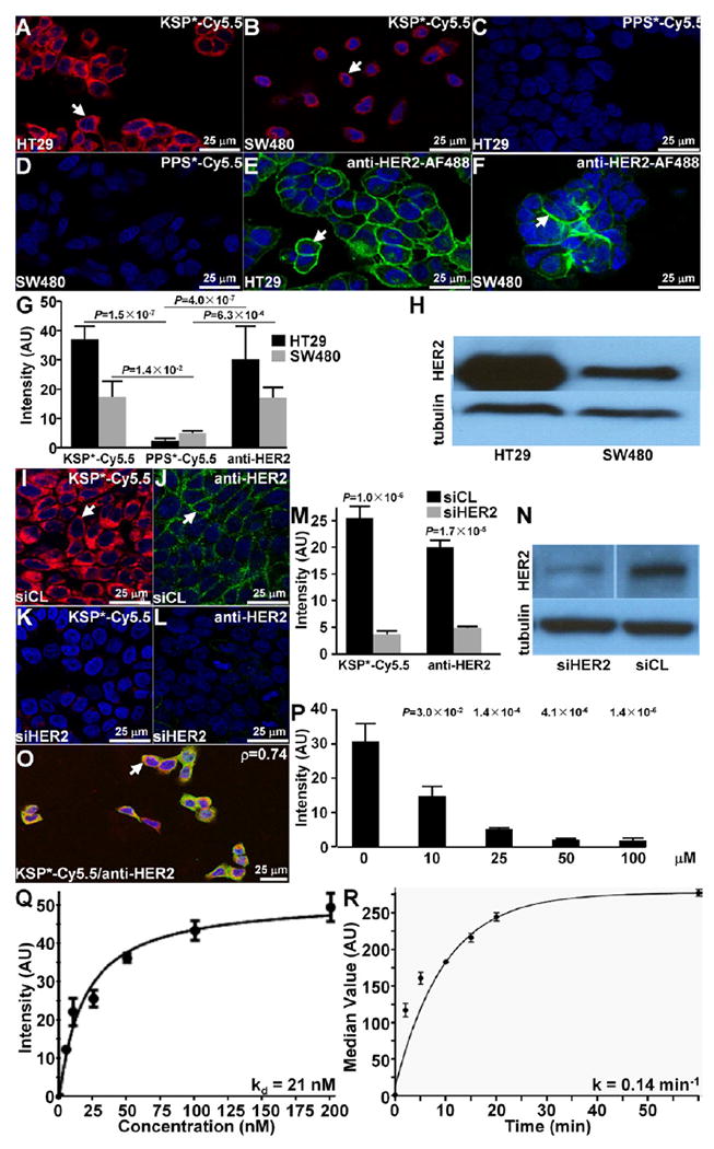
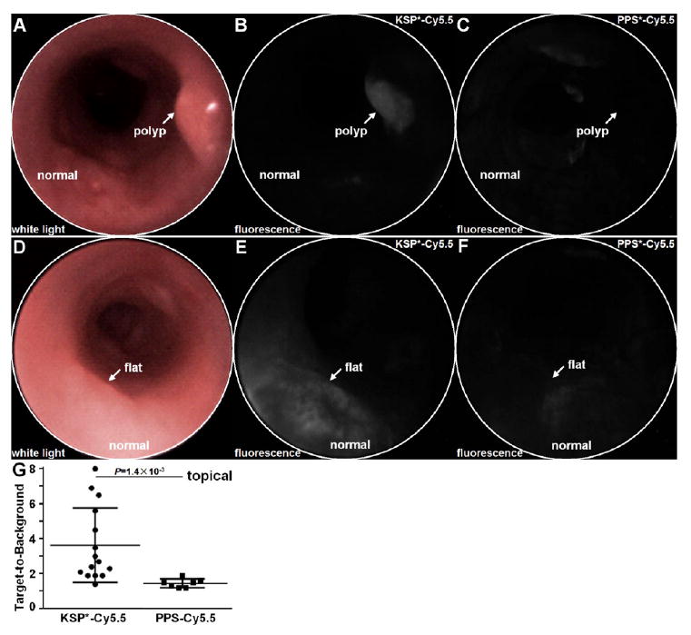
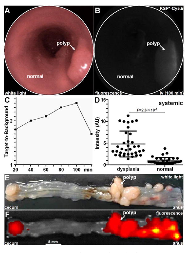
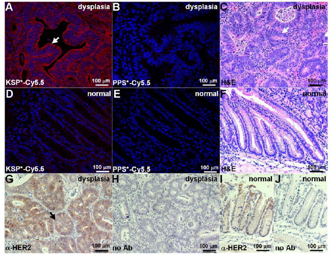
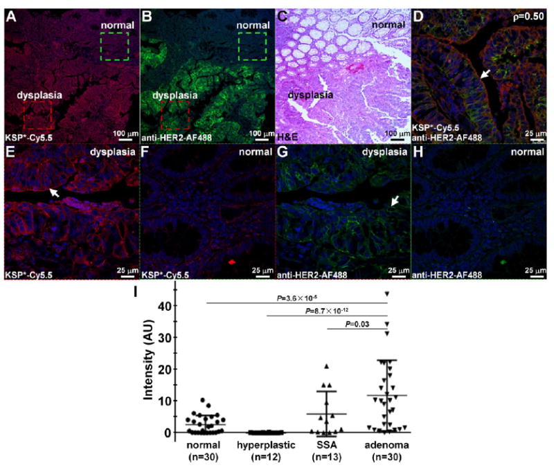
We report the development, characterization, and validation of a peptide specific for the extracellular domain of HER2. This probe chemistry was developed for molecular imaging by using a structural model to select an optimal combination of amino acids that maximize the likelihood for unique hydrophobic and hydrophilic interactions with HER2 domain 3. The sequence KSPNPRF was identified and conjugated with either FITC or Cy5.5 via a GGGSK linker using Fmoc-mediated solid-phase synthesis to demonstrate flexibility for this chemical structure to be labeled with different fluorophores. A scrambled sequence was developed for control by altering the conformationally rigid spacer and moving both hydrophobic and hydrophilic amino acids on the C-terminus. We validated peptide specificity for HER2 in knockdown and competition experiments using human colorectal cancer cells in vitro, and measured a binding affinity of kd = 21 nM and time constant of k = 0.14 min(-1) (7.14 min). We used this peptide with either topical or intravenous administration in a preclinical model of colorectal cancer to demonstrate specific uptake in spontaneous adenomas and to show feasibility for real time in vivo imaging with near-infrared fluorescence. We used this peptide in immunofluorescence studies of human proximal colon specimens to evaluate specificity for sessile serrated and sporadic adenomas. Improved visualization can be used endoscopically to guide tissue biopsy and detect premalignant lesions that would otherwise be missed. Our peptide design for specificity to HER2 is promising for clinical translation in molecular imaging methods for early cancer detection.
我们报告了一种针对HER2细胞外结构域的肽的研发、特性鉴定及验证。这种探针化学技术是为分子成像而开发的,通过使用结构模型来选择氨基酸的最佳组合,以最大化与HER2结构域3发生独特疏水和亲水相互作用的可能性。确定了序列KSPNPRF,并通过Fmoc介导的固相合成,经由GGGSK接头与FITC或Cy5.5偶联,以证明这种化学结构用不同荧光团标记的灵活性。通过改变构象刚性间隔区并移动C端的疏水和亲水氨基酸,开发了一个乱序序列作为对照。我们在体外使用人结肠癌细胞进行的敲低和竞争实验中验证了该肽对HER2的特异性,并测得结合亲和力kd = 21 nM,时间常数k = 0.14 min⁻¹(7.14分钟)。我们在结直肠癌的临床前模型中局部或静脉注射使用这种肽,以证明在自发性腺瘤中的特异性摄取,并展示近红外荧光实时体内成像的可行性。我们在人近端结肠标本的免疫荧光研究中使用这种肽,以评估对无蒂锯齿状腺瘤和散发性腺瘤的特异性。改善的可视化效果可在内窥镜检查中用于指导组织活检,并检测那些否则会被漏诊的癌前病变。我们针对HER2特异性的肽设计在用于早期癌症检测的分子成像方法的临床转化方面很有前景。