Department of Clinical Sciences, Intervention and Technology, Karolinska Institutet, 14186 Stockholm, Sweden.
Department of Clinical Sciences, Intervention and Technology, Karolinska Institutet, 14186 Stockholm, Sweden; Department of Clinical Neuroscience, Section for Ophthalmology and Vision, St. Erik Eye Hospital, Karolinska Institutet, 11282 Stockholm, Sweden.
Stem Cell Reports. 2016 Jan 12;6(1):9-17. doi: 10.1016/j.stemcr.2015.11.008. Epub 2015 Dec 24.
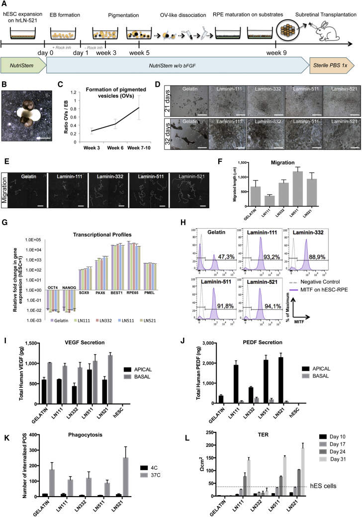
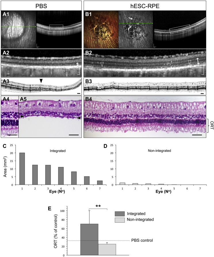
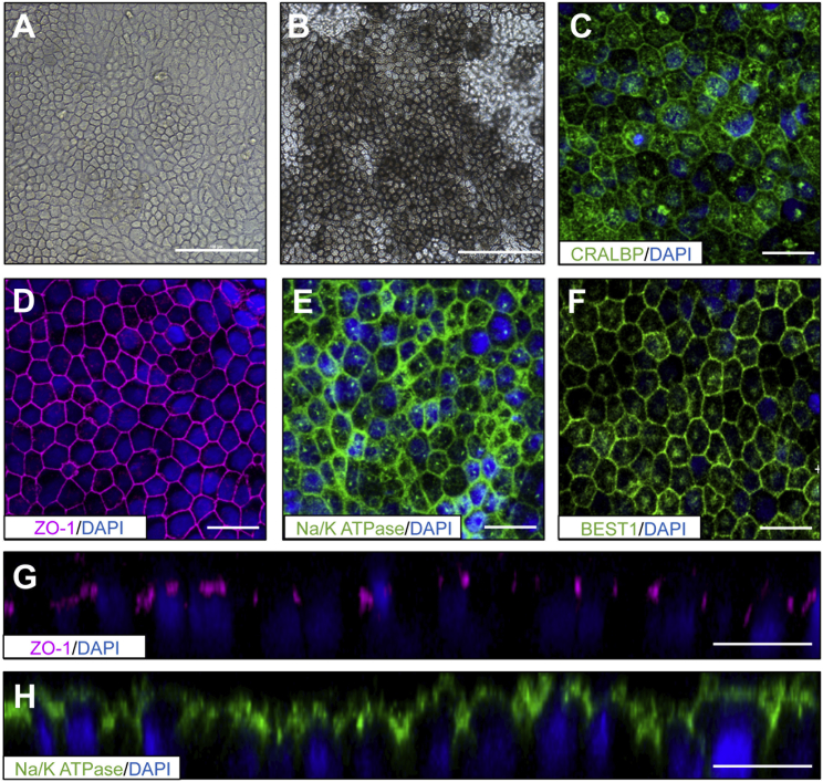
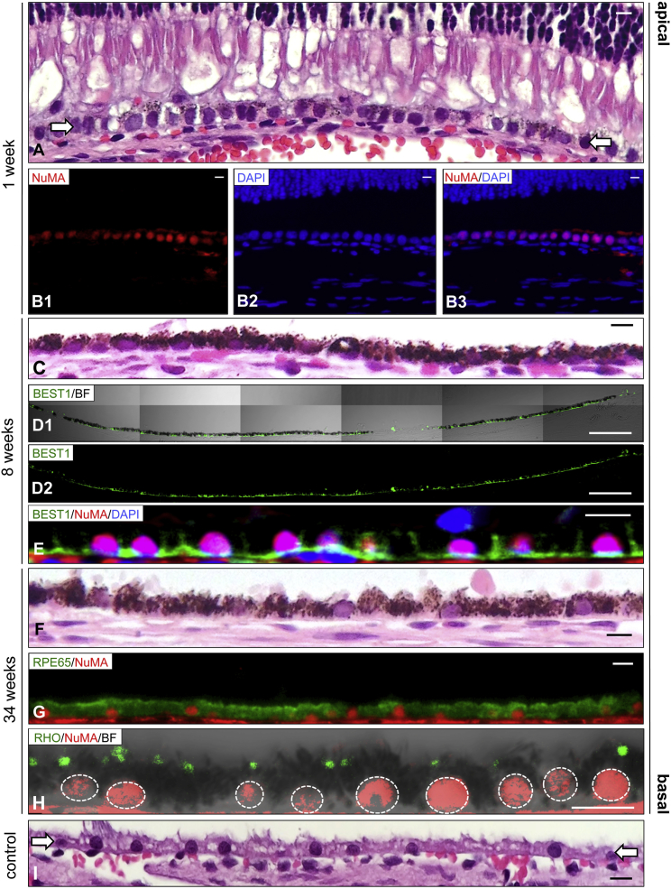
Human embryonic stem cell (hESC)-derived retinal pigment epithelial (RPE) cells could replace lost tissue in geographic atrophy (GA) but efficacy has yet to be demonstrated in a large-eyed model. Also, production of hESC-RPE has not yet been achieved in a xeno-free and defined manner, which is critical for clinical compliance and reduced immunogenicity. Here we describe an effective differentiation methodology using human laminin-521 matrix with xeno-free and defined medium. Differentiated cells exhibited characteristics of native RPE including morphology, pigmentation, marker expression, monolayer integrity, and polarization together with phagocytic activity. Furthermore, we established a large-eyed GA model that allowed in vivo imaging of hESC-RPE and host retina. Cells transplanted in suspension showed long-term integration and formed polarized monolayers exhibiting phagocytic and photoreceptor rescue capacity. We have developed a xeno-free and defined hESC-RPE differentiation method and present evidence of functional integration of clinically compliant hESC-RPE in a large-eyed disease model.
人胚胎干细胞(hESC)衍生的视网膜色素上皮(RPE)细胞可以替代地理萎缩(GA)中丢失的组织,但在大眼模型中尚未证明其疗效。此外,尚未以无动物和定义的方式生产 hESC-RPE,这对于临床合规性和降低免疫原性至关重要。在这里,我们描述了一种使用无动物和定义的培养基的有效的分化方法,使用人层粘连蛋白-521 基质。分化的细胞表现出具有天然 RPE 特征的形态、色素沉着、标志物表达、单层完整性和极化以及吞噬活性。此外,我们建立了一个大眼 GA 模型,允许对 hESC-RPE 和宿主视网膜进行体内成像。以悬浮液形式移植的细胞表现出长期整合,并形成具有吞噬和光感受器拯救能力的极化单层。我们已经开发了一种无动物和定义的 hESC-RPE 分化方法,并提供了在大眼疾病模型中具有临床合规性的 hESC-RPE 功能整合的证据。