Tennakoon Anusha H, Izawa Takeshi, Kuwamura Mitsuru, Yamate Jyoji
Laboratory of Veterinary Pathology, Life and Environmental Sciences, Osaka Prefecture University, Rinkuu Ourai Kita 1-58, Izumisano, Osaka 598-8531, Japan.
Teaching Hospital Peradeniya, Peradeniya 20400, Sri Lanka.
J Clin Med. 2015 Dec 30;5(1):4. doi: 10.3390/jcm5010004.
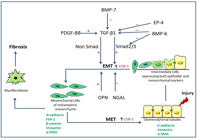
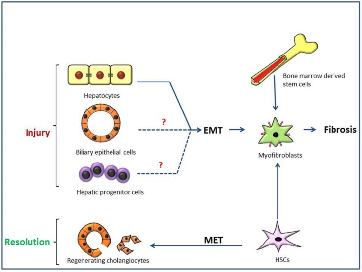
Epithelial to mesenchymal transition (EMT), particularly, type 2 EMT, is important in progressive renal and hepatic fibrosis. In this process, incompletely regenerated renal epithelia lose their epithelial characteristics and gain migratory mesenchymal qualities as myofibroblasts. In hepatic fibrosis (importantly, cirrhosis), the process also occurs in injured hepatocytes and hepatic progenitor cells (HPCs), as well as ductular reaction-related bile epithelia. Interestingly, the ductular reaction contributes partly to hepatocarcinogenesis of HPCs, and further, regenerating cholangiocytes after injury may be derived from hepatic stellate cells via mesenchymal to epithelia transition, a reverse phenomenon of type 2 EMT. Possible pathogenesis of type 2 EMT and its differences between renal and hepatic fibrosis are reviewed based on our experimental data.
上皮-间质转化(EMT),尤其是2型EMT,在进行性肾纤维化和肝纤维化中起重要作用。在此过程中,未完全再生的肾上皮细胞失去其上皮特征,并获得成肌纤维细胞的迁移性间质特性。在肝纤维化(重要的是肝硬化)中,该过程也发生在受损的肝细胞和肝祖细胞(HPC)以及与小胆管反应相关的胆管上皮细胞中。有趣的是,小胆管反应部分促成了HPC的肝癌发生,此外,损伤后再生的胆管细胞可能通过间质-上皮转化(2型EMT的反向现象)源自肝星状细胞。基于我们的实验数据,对2型EMT的可能发病机制及其在肾纤维化和肝纤维化之间的差异进行了综述。