Fabris Luca, Brivio Simone, Cadamuro Massimiliano, Strazzabosco Mario
Department of Molecular Medicine, University of Padua School of Medicine, Viale G. Colombo 3, 35131 Padua, Italy; Liver Center, Section of Digestive Diseases, Yale University, TAC Building, 333 Cedar Street, New Haven, CT 06520, USA.
School of Medicine and Surgery, University of Milan-Bicocca, Via Cadore 48, 20900 Monza, Italy.
Stem Cells Int. 2016;2016:2953727. doi: 10.1155/2016/2953727. Epub 2016 Jan 6.
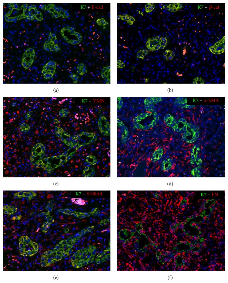
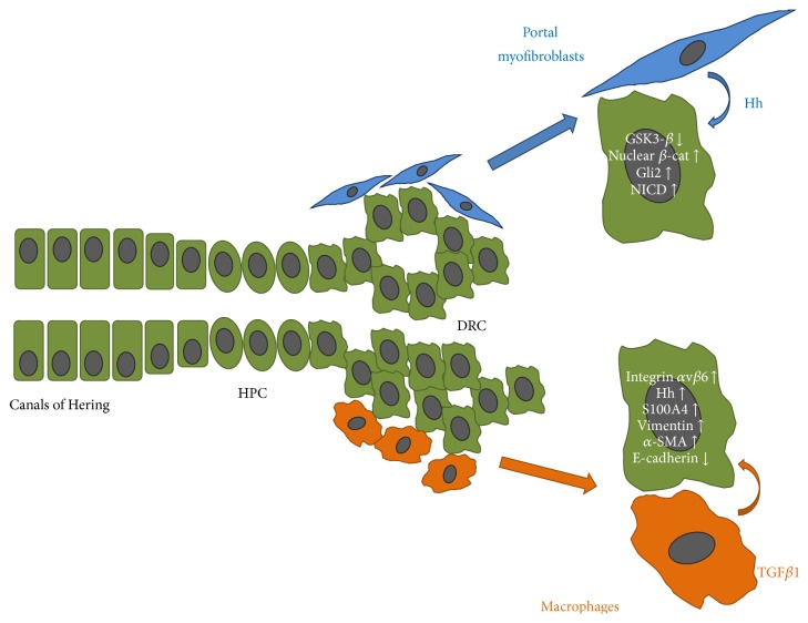
Whether liver epithelial cells contribute to the development of hepatic scarring by undergoing epithelial-to-mesenchymal transition (EMT) is a controversial issue. Herein, we revisit the concept of EMT in cholangiopathies, a group of severe hepatic disorders primarily targeting the bile duct epithelial cell (cholangiocyte), leading to progressive portal fibrosis, the main determinant of liver disease progression. Unfortunately, therapies able to halt this process are currently lacking. In cholangiopathies, fibrogenesis is part of ductular reaction, a reparative complex involving epithelial, mesenchymal, and inflammatory cells. Ductular reactive cells (DRC) are cholangiocytes derived from the activation of the hepatic progenitor cell compartment. These cells are arranged into irregular strings and express a "reactive" phenotype, which enables them to extensively crosstalk with the other components of ductular reaction. We will first discuss EMT in liver morphogenesis and then highlight how some of these developmental programs are partly reactivated in DRC. Evidence for "bona fide" EMT changes in cholangiocytes is lacking, but expression of some mesenchymal markers represents a fundamental repair mechanism in response to chronic biliary damage with potential harmful fibrogenetic effects. Understanding microenvironmental cues and signaling perturbations promoting these changes in DRC may help to identify potential targets for new antifibrotic therapies in cholangiopathies.
肝上皮细胞是否通过经历上皮-间质转化(EMT)促进肝纤维化的发展是一个有争议的问题。在此,我们重新审视胆管疾病中EMT的概念,胆管疾病是一组主要针对胆管上皮细胞(胆管细胞)的严重肝脏疾病,会导致进行性门静脉纤维化,这是肝病进展的主要决定因素。不幸的是,目前缺乏能够阻止这一过程的疗法。在胆管疾病中,纤维化是小胆管反应的一部分,小胆管反应是一种涉及上皮、间质和炎症细胞的修复复合体。小胆管反应性细胞(DRC)是源自肝祖细胞区室激活的胆管细胞。这些细胞排列成不规则的条索状,并表达一种“反应性”表型,这使它们能够与小胆管反应的其他成分广泛相互作用。我们将首先讨论肝形态发生中的EMT,然后强调这些发育程序中的一些如何在DRC中部分重新激活。胆管细胞中缺乏“真正的”EMT变化的证据,但一些间充质标志物的表达是对慢性胆道损伤的一种基本修复机制,可能具有有害的纤维化作用。了解促进DRC中这些变化的微环境线索和信号扰动,可能有助于确定胆管疾病新抗纤维化疗法的潜在靶点。