Zhang Liang, Trushin Sergey, Christensen Trace A, Bachmeier Benjamin V, Gateno Benjamin, Schroeder Andreas, Yao Jia, Itoh Kie, Sesaki Hiromi, Poon Wayne W, Gylys Karen H, Patterson Emily R, Parisi Joseph E, Diaz Brinton Roberta, Salisbury Jeffrey L, Trushina Eugenia
Department of Neurology, Mayo Clinic, 200 First St. SW, Rochester, MN 55905.
Electron Microscopy Core Facility, Mayo Clinic, 200 First St. SW, Rochester, MN 55905.
Sci Rep. 2016 Jan 5;6:18725. doi: 10.1038/srep18725.
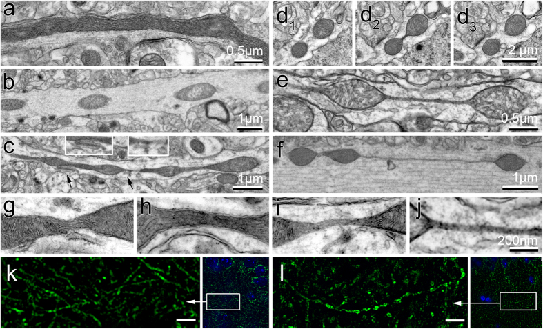
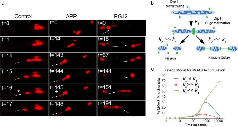
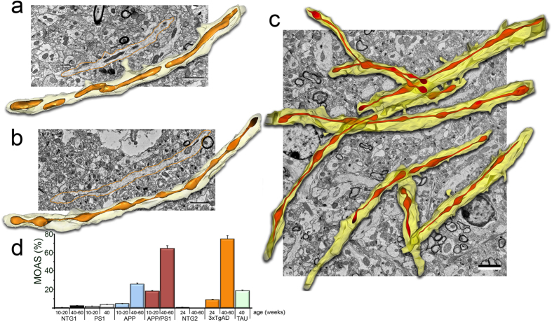
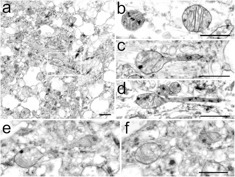
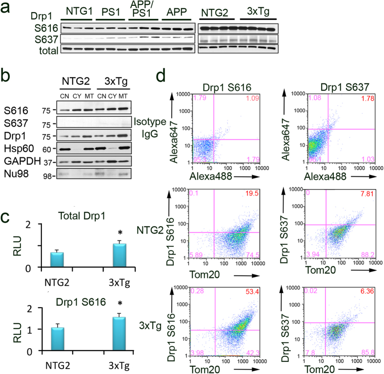
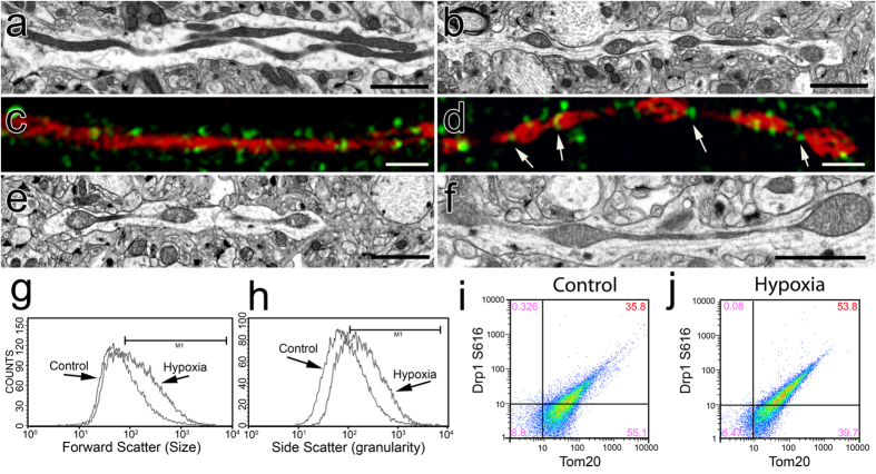
Altered brain metabolism is associated with progression of Alzheimer's Disease (AD). Mitochondria respond to bioenergetic changes by continuous fission and fusion. To account for three dimensional architecture of the brain tissue and organelles, we applied 3-dimensional electron microscopy (3D EM) reconstruction to visualize mitochondrial structure in the brain tissue from patients and mouse models of AD. We identified a previously unknown mitochondrial fission arrest phenotype that results in elongated interconnected organelles, "mitochondria-on-a-string" (MOAS). Our data suggest that MOAS formation may occur at the final stages of fission process and was not associated with altered translocation of activated dynamin related protein 1 (Drp1) to mitochondria but with reduced GTPase activity. Since MOAS formation was also observed in the brain tissue of wild-type mice in response to hypoxia or during chronological aging, fission arrest may represent fundamental compensatory adaptation to bioenergetic stress providing protection against mitophagy that may preserve residual mitochondrial function. The discovery of novel mitochondrial phenotype that occurs in the brain tissue in response to energetic stress accurately detected only using 3D EM reconstruction argues for a major role of mitochondrial dynamics in regulating neuronal survival.
大脑代谢改变与阿尔茨海默病(AD)的进展相关。线粒体通过持续的分裂和融合对生物能量变化做出反应。为了研究脑组织和细胞器的三维结构,我们应用三维电子显微镜(3D EM)重建技术来观察AD患者和小鼠模型脑组织中的线粒体结构。我们发现了一种以前未知的线粒体分裂停滞表型,其导致线粒体呈细长的相互连接状态,即“线状线粒体”(MOAS)。我们的数据表明,MOAS的形成可能发生在分裂过程的最后阶段,且与激活的动力相关蛋白1(Drp1)向线粒体的转位改变无关,而是与GTPase活性降低有关。由于在野生型小鼠的脑组织中,缺氧或自然衰老过程中也观察到了MOAS的形成,分裂停滞可能代表了对生物能量应激的一种基本代偿性适应,可防止线粒体自噬,从而可能保留残余的线粒体功能。仅通过3D EM重建才能准确检测到的、在脑组织中因能量应激而出现的新型线粒体表型的发现,表明线粒体动力学在调节神经元存活中起主要作用。