Travis Katherine E, Golden Neville H, Feldman Heidi M, Solomon Murray, Nguyen Jenny, Mezer Aviv, Yeatman Jason D, Dougherty Robert F
Division of Neonatal and Developmental Medicine, Department of Pediatrics, Stanford University School of Medicine, Palo Alto, CA 94303, USA.
Division of Adolescent Medicine, Department of Pediatrics, Stanford University School of Medicine, Palo Alto, CA 94303, USA.
Neuroimage Clin. 2015 Oct 23;9:648-59. doi: 10.1016/j.nicl.2015.10.008. eCollection 2015.
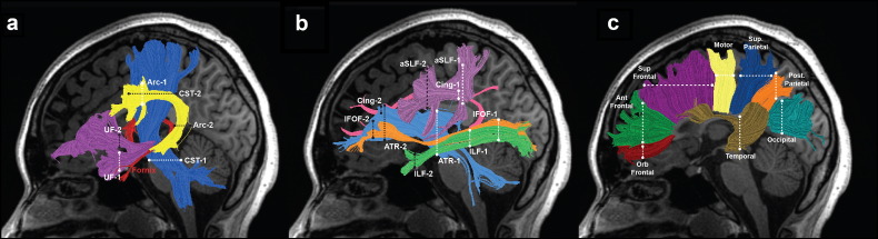
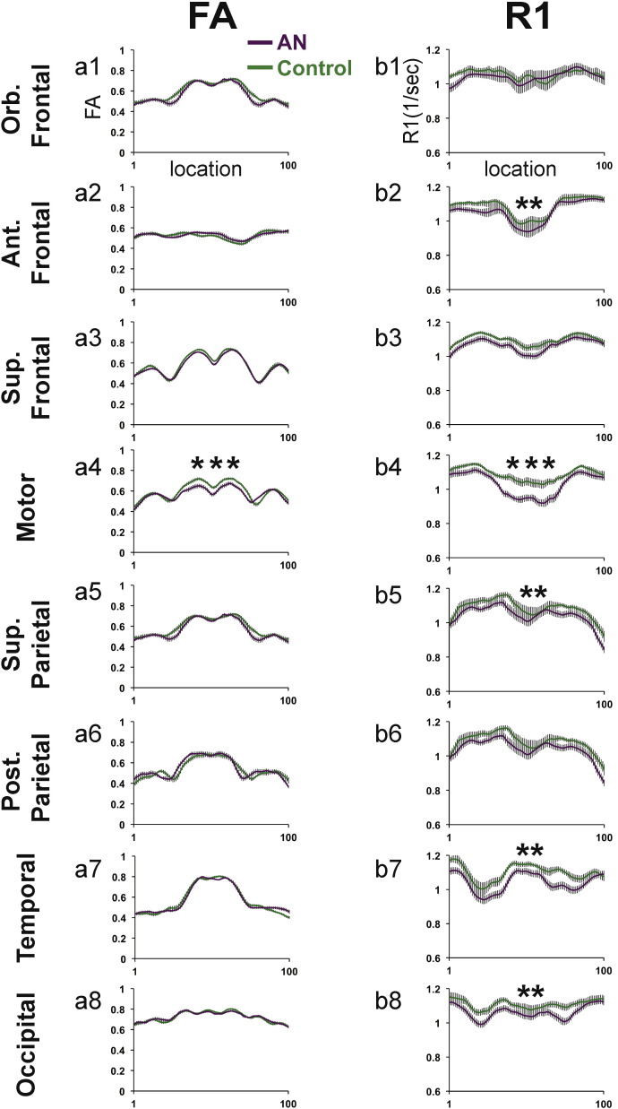
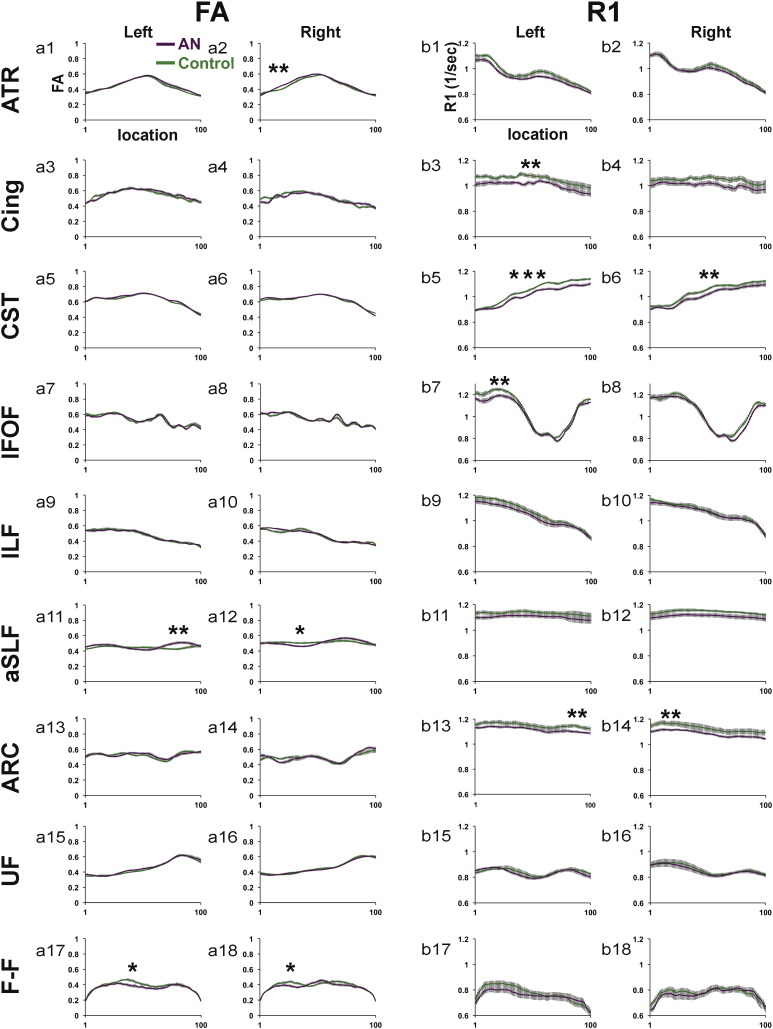
Anorexia nervosa (AN) is a serious eating disorder that typically emerges during adolescence and occurs most frequently in females. To date, very few studies have investigated the possible impact of AN on white matter tissue properties during adolescence, when white matter is still developing. The present study evaluated white matter tissue properties in adolescent girls with AN using diffusion MRI with tractography and T1 relaxometry to measure R1 (1/T1), an index of myelin content. Fifteen adolescent girls with AN (mean age = 16.6 years ± 1.4) were compared to fifteen age-matched girls with normal weight and eating behaviors (mean age = 17.1 years ± 1.3). We identified and segmented 9 bilateral cerebral tracts (18) and 8 callosal fiber tracts in each participant's brain (26 total). Tract profiles were generated by computing measures for fractional anisotropy (FA) and R1 along the trajectory of each tract. Compared to controls, FA in the AN group was significantly decreased in 4 of 26 white matter tracts and significantly increased in 2 of 26 white matter tracts. R1 was significantly decreased in the AN group compared to controls in 11 of 26 white matter tracts. Reduced FA in combination with reduced R1 suggests that the observed white matter differences in AN are likely due to reductions in myelin content. For the majority of tracts, group differences in FA and R1 did not occur within the same tract. The present findings have important implications for understanding the neurobiological factors underlying white matter changes associated with AN and invite further investigations examining associations between white matter properties and specific physiological, cognitive, social, or emotional functions affected in AN.
神经性厌食症(AN)是一种严重的饮食失调症,通常在青春期出现,且在女性中最为常见。迄今为止,很少有研究调查神经性厌食症在青春期对白质组织特性可能产生的影响,而青春期白质仍在发育。本研究使用扩散磁共振成像(MRI)结合纤维束成像和T1弛豫测量法来测量R1(1/T1,髓鞘含量指标),评估患有神经性厌食症的青春期女孩的白质组织特性。将15名患有神经性厌食症的青春期女孩(平均年龄 = 16.6岁±1.4)与15名年龄匹配、体重和饮食行为正常的女孩(平均年龄 = 17.1岁±1.3)进行比较。我们在每个参与者的大脑中识别并分割出9条双侧脑纤维束(共18条)和8条胼胝体纤维束(共26条)。通过计算每条纤维束轨迹上的分数各向异性(FA)和R1测量值来生成纤维束剖面图。与对照组相比,神经性厌食症组在26条白质纤维束中有4条的FA显著降低,有2条的FA显著升高。在26条白质纤维束中,神经性厌食症组的R1与对照组相比有11条显著降低。FA降低与R1降低相结合表明,在神经性厌食症中观察到的白质差异可能是由于髓鞘含量减少所致。对于大多数纤维束而言,FA和R1的组间差异并非出现在同一条纤维束中。本研究结果对于理解与神经性厌食症相关的白质变化背后的神经生物学因素具有重要意义,并促使进一步研究白质特性与神经性厌食症中受影响的特定生理、认知、社会或情感功能之间的关联。