Yin Bing, Li Wenhua, Cui Yanfen, Chu Caiting, Ding Ming, Chen Jian, Zhang Ping, Wu Xiangru
Department of Radiology, Xinhua Hospital affiliated to Shanghai Jiao Tong University School of Medicine, Shanghai, 200092, China.
Department of Obstetrics and Gynecology, Xinhua Hospital affiliated to Shanghai Jiao Tong University School of Medicine, Shanghai, 200092, China.
World J Surg Oncol. 2016 Jan 8;14(1):5. doi: 10.1186/s12957-015-0760-x.
Our study aims to determine the value of diffusion-weighted imaging (DWI) combined with conventional magnetic resonance imaging (MRI) in the diagnosis of thecomas/fibrothecomas and their differential diagnosis with malignant pelvic solid tumors.
In total, 36 thecomas/fibrothecomas and 40 malignant pelvic solid tumors were included in our study. All patients underwent 1.5 T conventional MRI and DWI examinations except one patient with a fibrothecoma in whom DWI examination was not performed. The clinical features and characteristics of conventional MRI and DWI of these two groups were analyzed. Apparent diffusion coefficient (ADC) values were measured and compared between groups. Univariate analysis, multivariate logistic regression analysis, and the receiver operating characteristic curve were used for statistical analysis.
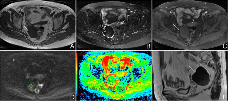
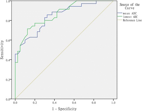
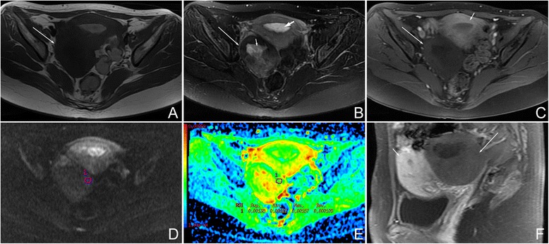
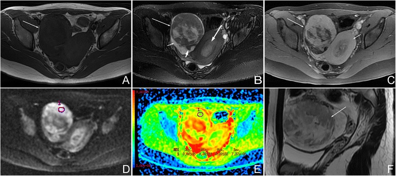
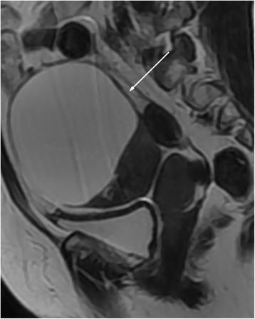
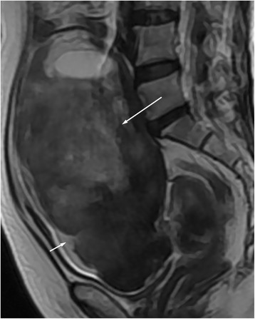
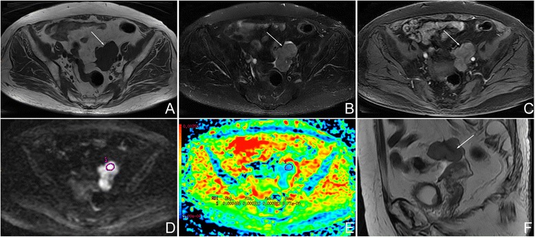
All the thecomas/fibrothecomas showed isointensity on T1 weighted imaging (T1WI) and 77.8% (28/36) lesions showed hypo- to isointensity on T2 weighted imaging (T2WI). After administration of contrast medium, 94.4% (34/36) tumors appeared as minor to mild enhancement. On DWI, they showed a diversity of low to very high signal intensity. All malignant pelvic masses manifested as hyperintensity on T2WI and 87.5% (35/40) tumors showed very high signal (grade 3) on DWI. Higher area under the curve (AUC) and specificity could be achieved by using the lowest ADC value than the mean ADC value. Multivariate logistic regression analysis showed that shape, signal intensity on T2WI, capsule, and the lowest ADC value were the important indicators in discriminating thecomas/fibrothecomas from malignant pelvic solid tumors.
The combination of DWI and conventional MRI is of great value in the diagnosis of thecomas/fibrothecomas and their differential diagnosis with malignant pelvic solid tumors.
本研究旨在确定弥散加权成像(DWI)联合传统磁共振成像(MRI)在诊断卵泡膜瘤/纤维卵泡膜瘤及其与盆腔恶性实性肿瘤鉴别诊断中的价值。
本研究共纳入36例卵泡膜瘤/纤维卵泡膜瘤和40例盆腔恶性实性肿瘤。除1例纤维卵泡膜瘤患者未行DWI检查外,所有患者均接受了1.5T传统MRI和DWI检查。分析两组患者的临床特征以及传统MRI和DWI的特点。测量并比较两组的表观扩散系数(ADC)值。采用单因素分析、多因素logistic回归分析及受试者工作特征曲线进行统计学分析。
所有卵泡膜瘤/纤维卵泡膜瘤在T1加权成像(T1WI)上呈等信号,77.8%(28/36)的病灶在T2加权成像(T2WI)上呈低至等信号。注射造影剂后,94.4%(34/36)的肿瘤呈轻度至中度强化。在DWI上,它们表现出从低到非常高信号强度的多样性。所有盆腔恶性肿块在T2WI上均表现为高信号,87.5%(35/40)的肿瘤在DWI上表现为非常高信号(3级)。使用最低ADC值比平均ADC值可获得更高的曲线下面积(AUC)和特异性。多因素logistic回归分析显示,形态、T2WI信号强度、包膜及最低ADC值是鉴别卵泡膜瘤/纤维卵泡膜瘤与盆腔恶性实性肿瘤的重要指标。
DWI与传统MRI相结合在卵泡膜瘤/纤维卵泡膜瘤的诊断及其与盆腔恶性实性肿瘤的鉴别诊断中具有重要价值。