Tyagi Neelam, Riaz Nadeem, Hunt Margie, Wengler Kenneth, Hatzoglou Vaios, Young Robert, Mechalakos James, Lee Nancy
Department of Medical Physics, Memorial Sloan-Kettering Cancer Center, 1275 York Avenue, New York, New York 10065.
Department of Radiation Oncology, Memorial Sloan-Kettering Cancer Center, 1275 York Avenue, New York, New York 10065.
Med Phys. 2016 Jan;43(1):137. doi: 10.1118/1.4937791.
Patients with cancers of oropharynx have a favorable prognosis and are an ideal candidate for adaptive therapy. A replan to improve coverage or escalate/de-escalate dose based on morphological information alone may not be adequate as the grossly involved lymph nodes (LNs) of a subset of these patients tend to become cystic and often do not regress. Functional adaptation may be a better approach when considering replanning for these patients. The purpose of this study was to evaluate the weekly trends in treatment related morphological and physiological changes for these LNs using diffusion-weighted MRI (DW-MRI) and evaluate its implications for adaptive replanning.
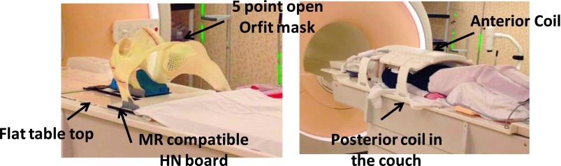
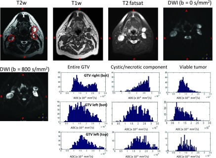
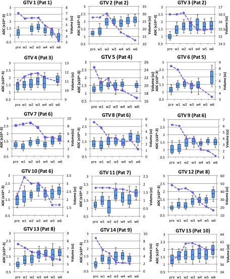
Ten patients with histologically proven oropharynx HNSCC undergoing concurrent chemoradiation were analyzed in this study. MR imaging protocol included axial T1w, T2w, and DW-MRI using a 3 T Philips MR scanner. The patients were scanned weekly in radiation treatment planning position using a 16 element phased-array anterior coil and a 44 element posterior coil. A total of 65 DWI and T2w scans were analyzed. DWI was performed using an optimized single-shot echo planar imaging sequence (TR/TE = 5000/65 ms, slice thickness = 5 mm; slices = 28; b values = 0 and 800 s/mm(2)). Quantification of the DW-MRI images was performed by calculating the apparent diffusion coefficient (ADC). T2w and DWI scans were imported into the Eclipse treatment planning system and gross tumor volumes (GTVs) corresponding to grossly involved LNs were contoured on each axial slice by physician experts. An attempt was made to remove any cystic or necrotic components so that the ADC analysis was of viable tumor only. A pixel-by-pixel fit of signal intensities within the GTVs was performed assuming monoexponential behavior. From each GTV histogram mean, median, standard deviation, skewness, and kurtosis were calculated. Absolute and percent change in weekly ADC histogram parameters and percent change in T2w GTV were also calculated.
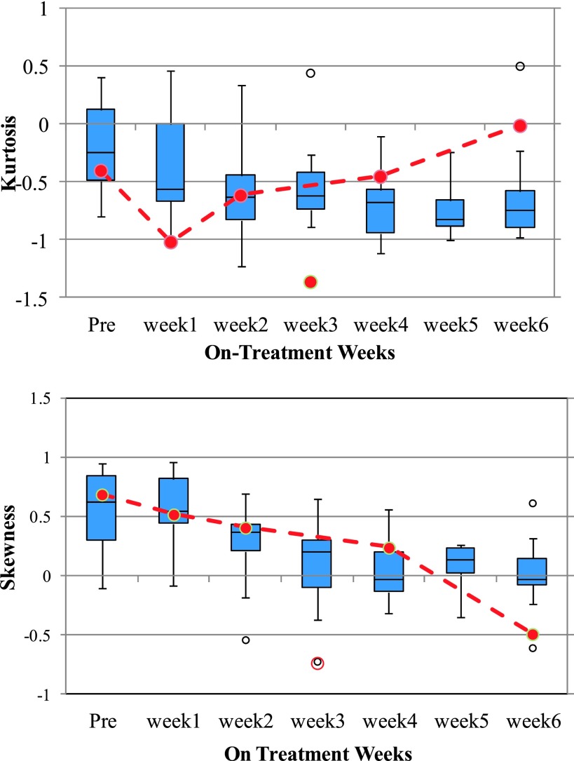
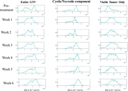
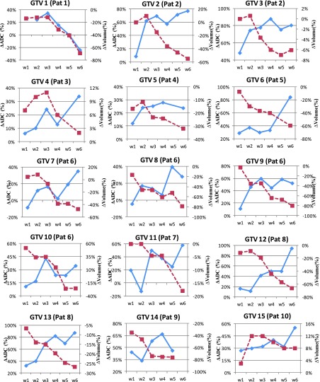
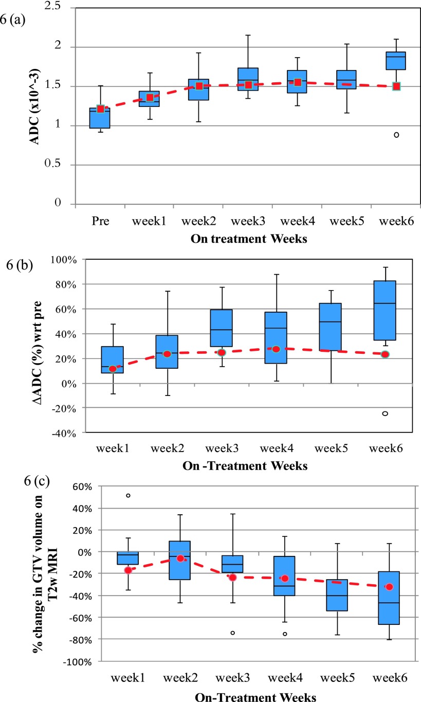
For all nodes, an immediate change in ADC was observed during first 2-3 weeks after which ADC values either continued to increase or plateaued. A few nodal volumes had a slightly decreased ADC value during later weeks. Percent increase in median ADC from weeks 1 to 6 with respect to baseline was 14%, 25%, 41%, 42%, 45%, and 58%. The corresponding change in median T2 volumes was 8%, 10%, 16%, 22%, 40%, and 42%, respectively. The ADC distribution of the viable tumors was initially highly kurtotic; however, the kurtosis decreased as treatment progressed. The ADC distribution also showed a higher degree of skewness in the first 2 weeks, progressively becoming less skewed as treatment progressed so as to slowly approach a more symmetric distribution.
Physiological changes in LNs represented by changes in ADC evaluated using DW-MRI are evident sooner than the morphological changes calculated from T2w MRI. The decisions for adaptive replanning may need to be individualized and should be based primarily on tumor functional information. The authors' data also suggest that for many patients, week 3 maybe the optimal time to intervene and replan. Larger studies are needed to confirm their findings.
口咽癌患者预后良好,是适应性治疗的理想候选者。仅基于形态学信息重新规划以改善覆盖范围或增加/减少剂量可能并不充分,因为这些患者中的一部分其受累肿大淋巴结往往会变成囊性,且通常不会消退。在考虑为这些患者重新规划时,功能适应性可能是一种更好的方法。本研究的目的是使用扩散加权磁共振成像(DW-MRI)评估这些淋巴结与治疗相关的形态学和生理学变化的每周趋势,并评估其对适应性重新规划的影响。
本研究分析了10例经组织学证实为口咽癌的头颈部鳞状细胞癌患者,这些患者正在接受同步放化疗。磁共振成像方案包括使用3T飞利浦磁共振扫描仪进行的轴位T1加权、T2加权和DW-MRI检查。患者每周在放射治疗计划体位下使用16元素相控阵前线圈和44元素后线圈进行扫描。共分析了65次DWI和T2加权扫描。DWI使用优化的单次激发回波平面成像序列(TR/TE = 5000/65 ms,层厚 = 5 mm;层数 = 28;b值 = 0和800 s/mm²)进行。通过计算表观扩散系数(ADC)对DW-MRI图像进行定量分析。将T2加权和DWI扫描图像导入Eclipse治疗计划系统,由医生专家在每个轴位切片上勾勒出与受累肿大淋巴结相对应的大体肿瘤体积(GTV)。试图去除任何囊性或坏死成分,以便ADC分析仅针对存活肿瘤。假设信号强度呈单指数行为,对GTV内的信号强度进行逐像素拟合。从每个GTV直方图中计算均值、中位数、标准差、偏度和峰度。还计算了每周ADC直方图参数的绝对变化和百分比变化以及T2加权GTV的百分比变化。
对于所有淋巴结,在最初的2 - 3周内观察到ADC立即发生变化,此后ADC值要么继续增加,要么趋于平稳。在随后的几周内,少数淋巴结体积的ADC值略有下降。第1至6周相对于基线,ADC中位数的百分比增加分别为14%、25%、41%、42%、45%和58%。相应的T2体积中位数变化分别为8%、10%、16%、22%、40%和42%。存活肿瘤的ADC分布最初具有高度峰度;然而,随着治疗进展,峰度降低。ADC分布在最初2周也显示出较高程度的偏度,随着治疗进展逐渐变得不那么偏斜,从而缓慢接近更对称的分布。
使用DW-MRI评估的以ADC变化表示的淋巴结生理学变化比从T2加权MRI计算的形态学变化更明显。适应性重新规划的决策可能需要个体化,并且应主要基于肿瘤功能信息。作者的数据还表明,对于许多患者,第3周可能是进行干预和重新规划的最佳时间。需要更大规模的研究来证实他们的发现。