Yudistiro Ryan, Arisaka Yukiko, Tokue Azusa, Nakajima Takahito
Department of Diagnostic Radiology and Nuclear Medicine, Gunma University Graduate School of Medicine, 3-39-22 Showa, Maebashi, 371-8511, Gunma, Japan.
Department of Diagnostic Radiology and Nuclear Medicine, Gunma University Hospital, Gunma, Japan.
BMC Med Imaging. 2016 Jan 8;16:1. doi: 10.1186/s12880-015-0104-x.
Sarcoidosis-lymphoma syndrome (SLS) is a rare disease in which both entities coexist. We aimed to study the role of (18)F-fluorodeoxyglucose (FDG) and L-[3-(18)F] α-methyltyrosine (FAMT) positron emission tomography (PET)/computed tomography (CT) in differentiating between these two lesions.
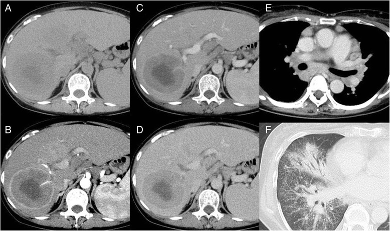
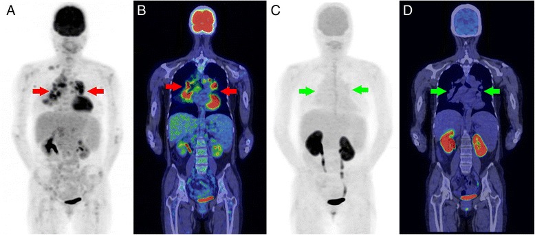
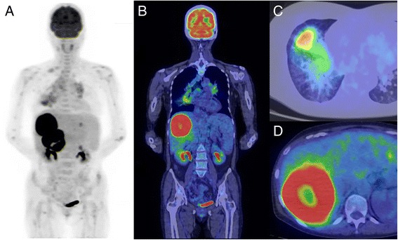
A 54-year-old female with large liver tumors was referred to our Nuclear Medicine Department for staging using FDG PET/CT. She had a history of primary biliary cirrhosis (PBC) for 15 years and developed lung and mediastinal sarcoidosis 1 year before the liver tumors were noted. Abdominal dynamic CT revealed two well-circumscribed, peripherally-enhancing, low-density masses in the right lobe of the liver with intensive ring-form FDG uptakes at maximum standard uptake values (SUVmax) of 18.3 and 19.5, respectively. In the arterial phase, a hepatic artery was seen penetrating the tumor, a phenomenon known as "angiogram sign". Chest PET/CT findings showed irregular thickening of the bronchovascular bundles, central peribronchial shaggy consolidations in the right middle and lower lobes (SUVmax, 4.6), and mediastinal and hilar lymphadenopathies (SUVmax, 2.7). After assessment, chemotherapy with rituximab, cyclophosphamide, doxorubicin hydrochloride, vincristine sulfate, and prednisone (R-CHOP) was administered for eight cycles. Follow-up imaging studies using FDG and FAMT PET/CT were performed 3 months after the last cycle of chemotherapy, which showed that the two highly FDG-avid tumors in the liver had disappeared. However, faint FDG uptake persisted in the lung consolidations (SUVmax, 6.3), and FDG uptake for the mediastinal lymphadenopathies increased (SUVmax of 5.8). In contrast, there was no significant uptake of FAMT in the liver, as well as in the lungs and the bilateral mediastinal lymphadenopathies. These discrepant uptakes between FDG and FAMT were compatible with sarcoidosis.
Combination of FDG and FAMT in PET/CT studies may play an important role in the management of SLS patients, especially in differentiating between sarcoidosis and lymphoma lesions.
结节病-淋巴瘤综合征(SLS)是一种罕见疾病,其中这两种病症同时存在。我们旨在研究18F-氟脱氧葡萄糖(FDG)和L-[3-(18)F]α-甲基酪氨酸(FAMT)正电子发射断层扫描(PET)/计算机断层扫描(CT)在鉴别这两种病变中的作用。
一名54岁患有肝脏巨大肿瘤的女性被转诊至我们核医学科,使用FDG PET/CT进行分期。她有原发性胆汁性肝硬化(PBC)病史15年,在发现肝脏肿瘤前1年出现肺部和纵隔结节病。腹部动态CT显示肝脏右叶有两个边界清晰、周边强化的低密度肿块,FDG摄取呈密集环形,最大标准摄取值(SUVmax)分别为18.3和19.5。在动脉期,可见肝动脉穿入肿瘤,此现象称为“血管造影征”。胸部PET/CT检查结果显示支气管血管束不规则增厚,右中叶和下叶中央支气管周围有模糊的实变影(SUVmax,4.6),以及纵隔和肺门淋巴结肿大(SUVmax,2.7)。评估后,给予利妥昔单抗、环磷酰胺、盐酸多柔比星、硫酸长春新碱和泼尼松(R-CHOP)化疗8个周期。在最后一个化疗周期后3个月进行了使用FDG和FAMT PET/CT的随访影像学检查,结果显示肝脏中两个高度摄取FDG的肿瘤消失。然而,肺部实变影中仍有微弱的FDG摄取(SUVmax,6.3),纵隔淋巴结肿大的FDG摄取增加(SUVmax为5.8)。相比之下,FAMT在肝脏以及肺部和双侧纵隔淋巴结肿大中均无明显摄取。FDG和FAMT之间这些不同的摄取情况与结节病相符。
PET/CT检查中FDG和FAMT的联合应用可能在SLS患者的管理中发挥重要作用,尤其是在鉴别结节病和淋巴瘤病变方面。