KleinJan Gijs H, van den Berg Nynke S, de Jong Jeroen, Wit Esther M, Thygessen Helene, Vegt Erik, van der Poel Henk G, van Leeuwen Fijs W B
Interventional Molecular Imaging Laboratory, Department of Radiology, Leiden University Medical Hospital, Albinusdreef 2, 2300RC, Leiden, The Netherlands.
Department of Nuclear Medicine, The Netherlands Cancer Institute - Antoni van Leeuwenhoek Hospital, Plesmanlaan 121, 1066CX, Amsterdam, The Netherlands.
Eur J Nucl Med Mol Imaging. 2016 Jul;43(7):1278-87. doi: 10.1007/s00259-015-3292-2. Epub 2016 Jan 15.
Radical prostatectomy and complementary extended pelvic lymph node dissection (ePLND) of sentinel lymph nodes (SNs) and non-sentinel lymph nodes (LNs) at risk of containing metastases are increasingly being performed using high-tech robot-assisted approaches. Although this technological evolution has clear advantages, the physical nature of robotic systems limits the integrated use of routine radioguided surgery technologies. Hence, engineering effort in robotics are focused on the integration of fluorescence guidance technologies. Using the hybrid SN tracer indocyanine green-(99m)Tc-nanocolloid (radioactive and fluorescent), for the first time in combination with a robot-integrated laparoscope, we investigated whether the robot-assisted approach affects the accuracy of fluorescence detection of SNs identified preoperatively using nuclear medicine.
The study included 55 patients (Briganti nomogram-based risk >5 % on LN metastases) scheduled for robot-assisted radical prostatectomy, SN biopsy and ePLND. Following indocyanine green-(99m)Tc-nanocolloid injection, preoperative nuclear imaging (lymphoscintigraphy and SPECT/CT) was used to locate the SN(s). The fluorescence laparoscope was used intraoperatively to identify the SN(s) with standard fluorescence settings (in 50 patients) and with customized settings (in 5 patients). The number and location of the SNs, the radioactive, fluorescence (both in vivo and ex vivo) and tumour status of the resected SNs/LNs, and postoperative complications were recorded and analysed.
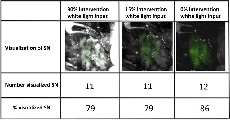
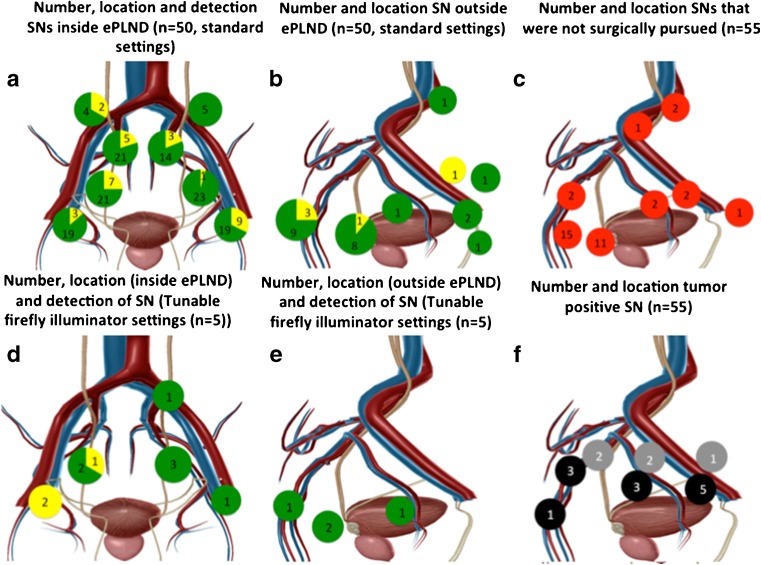
Combined, preoperative lymphoscintigraphy and SPECT/CT imaging identified 212 SNs (median 4 per patient). Intraoperative fluorescence imaging using standard fluorescence settings visualized 80.4 % (148/184 SNs; 50 patients; ex vivo 97.8 %). This increased to 85.7 % (12/14 SNs; 5 patients; ex vivo 100 %) with customized fluorescence settings. SPECT/CT images provided guidance towards the residual SNs. Ex vivo all removed SNs were radioactive. SNs were tumour-positive in 25.4 % of patients (14/55; false-negative rate 7 %, 1/14 patients). In ten patients, the SN was the only tumour-positive LN. Surgical complications were minimal.
Directly linking 3D preoperative nuclear imaging information on SNs to a robot-integrated fluorescence laparoscope improved the surgeon's use of the technology and did not influence the sensitivity or morbidity of the procedure. To our surprise, however, the detection rates with the current fluorescence camera did not improve.
根治性前列腺切除术以及对有转移风险的前哨淋巴结(SNs)和非前哨淋巴结(LNs)进行补充性扩大盆腔淋巴结清扫术(ePLND)越来越多地采用高科技机器人辅助方法进行。尽管这种技术进步具有明显优势,但机器人系统的物理特性限制了常规放射性引导手术技术的综合应用。因此,机器人技术的工程努力集中在荧光引导技术的集成上。我们首次将混合SN示踪剂吲哚菁绿 -(99m)Tc -纳米胶体(放射性和荧光性)与机器人集成腹腔镜相结合,研究机器人辅助方法是否会影响术前使用核医学识别的SNs荧光检测的准确性。
该研究纳入了55例计划进行机器人辅助根治性前列腺切除术、SN活检和ePLND的患者(基于Briganti列线图的LN转移风险>5%)。注射吲哚菁绿 -(99m)Tc -纳米胶体后,术前核成像(淋巴闪烁显像和SPECT/CT)用于定位SNs。术中使用荧光腹腔镜以标准荧光设置(50例患者)和定制设置(5例患者)识别SNs。记录并分析SNs的数量和位置、切除的SNs/LNs的放射性、荧光(体内和体外)以及肿瘤状态,以及术后并发症。
术前淋巴闪烁显像和SPECT/CT成像联合识别出212个SNs(每位患者中位数为4个)。使用标准荧光设置的术中荧光成像可视化了80.4%(148/184个SNs;50例患者;体外为97.8%)。定制荧光设置后这一比例增至85.7%(12/14个SNs;5例患者;体外为100%)。SPECT/CT图像为剩余的SNs提供了引导。体外所有切除的SNs均有放射性。25.4%的患者(14/55;假阴性率7%,1/14例患者)的SNs为肿瘤阳性。在10例患者中,SN是唯一的肿瘤阳性LN。手术并发症极少。
将术前关于SNs的3D核成像信息直接与机器人集成荧光腹腔镜相连接,改善了外科医生对该技术的使用,且不影响手术的敏感性或发病率。然而,令我们惊讶的是,当前荧光相机的检测率并未提高。