• 文献检索
  • 文档翻译
  • 深度研究
  • 学术资讯
  • Suppr Zotero 插件Zotero 插件
  • 邀请有礼
  • 套餐&价格
  • 历史记录
应用&插件
Suppr Zotero 插件Zotero 插件浏览器插件Mac 客户端Windows 客户端微信小程序
定价
高级版会员购买积分包购买API积分包
服务
文献检索文档翻译深度研究API 文档MCP 服务
关于我们
关于 Suppr公司介绍联系我们用户协议隐私条款
关注我们

Suppr 超能文献

核心技术专利:CN118964589B侵权必究
粤ICP备2023148730 号-1Suppr @ 2026

文献检索

告别复杂PubMed语法,用中文像聊天一样搜索,搜遍4000万医学文献。AI智能推荐,让科研检索更轻松。

立即免费搜索

文件翻译

保留排版,准确专业,支持PDF/Word/PPT等文件格式,支持 12+语言互译。

免费翻译文档

深度研究

AI帮你快速写综述,25分钟生成高质量综述,智能提取关键信息,辅助科研写作。

立即免费体验

细胞凋亡的荧光寿命成像

Fluorescence Lifetime Imaging of Apoptosis.

作者信息

Xiao Annie, Gibbons Anne E, Luker Kathryn E, Luker Gary D

机构信息

Center for Molecular Imaging, Department of Radiology, University of Michigan, Ann Arbor, MI, USA.

Departments of Biomedical Engineering and Microbiology and Immunology, University of Michigan, Ann Arbor, MI, USA.

出版信息

Tomography. 2015 Dec;1(2):115-124. doi: 10.18383/j.tom.2015.00163.

DOI:10.18383/j.tom.2015.00163
PMID:26771007
原文链接:https://pmc.ncbi.nlm.nih.gov/articles/PMC4710058/
Abstract

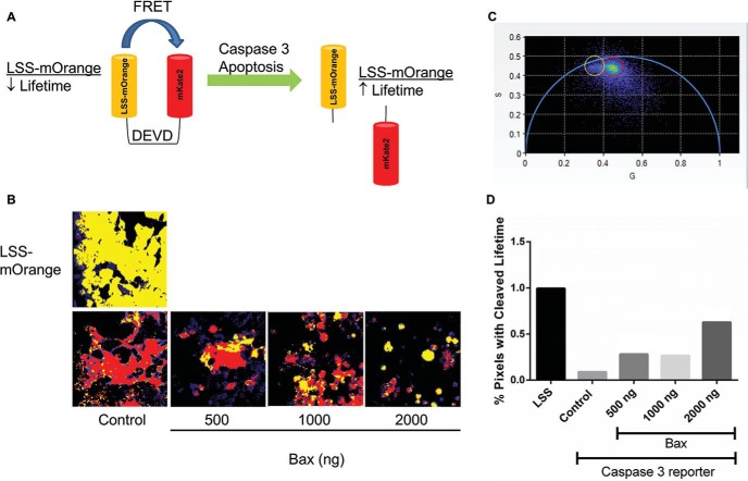
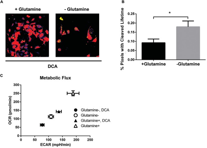
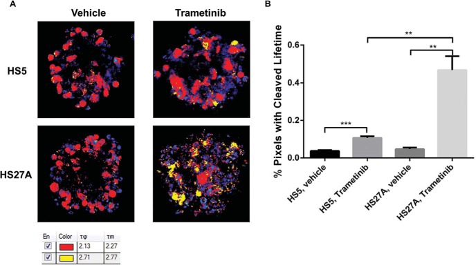
Genetically-encoded fluorescence resonance energy transfer (FRET) reporters are powerful tools to analyze cell signaling and function at single cell resolution in standard two-dimensional cell cultures, but these reporters rarely have been applied to three-dimensional environments. FRET interactions between donor and acceptor molecules typically are determined by changes in relative fluorescence intensities, but wavelength-dependent differences in absorption of light complicate this analysis method in three-dimensional settings. Here we report fluorescence lifetime imaging microscopy (FLIM) with phasor analysis, a method that displays fluorescence lifetimes on a pixel-wise basis in real time, to quantify apoptosis in breast cancer cells stably expressing a genetically encoded FRET reporter. This microscopic imaging technology allowed us to identify treatment-induced apoptosis in single breast cancer cells in environments ranging from two-dimensional cell culture, spheroids with cancer and bone marrow stromal cells, and living mice with orthotopic human breast cancer xenografts. Using this imaging strategy, we showed that combined metabolic therapy targeting glycolysis and glutamine pathways significantly reduced overall breast cancer metabolism and induced apoptosis. We also determined that distinct subpopulations of bone marrow stromal cells control resistance of breast cancer cells to chemotherapy, suggesting heterogeneity of treatment responses of malignant cells in different bone marrow niches. Overall, this study establishes FLIM with phasor analysis as an imaging tool for apoptosis in cell-based assays and living mice, enabling real-time, cellular-level assessment of treatment efficacy and heterogeneity.

摘要

基因编码的荧光共振能量转移(FRET)报告分子是在标准二维细胞培养中以单细胞分辨率分析细胞信号传导和功能的强大工具,但这些报告分子很少应用于三维环境。供体和受体分子之间的FRET相互作用通常由相对荧光强度的变化来确定,但光吸收的波长依赖性差异使这种分析方法在三维环境中变得复杂。在这里,我们报告了带有相量分析的荧光寿命成像显微镜(FLIM),这是一种实时逐像素显示荧光寿命的方法,用于量化稳定表达基因编码FRET报告分子的乳腺癌细胞中的凋亡。这种显微成像技术使我们能够在从二维细胞培养、含有癌细胞和骨髓基质细胞的球体以及患有原位人乳腺癌异种移植的活体小鼠等不同环境中识别单个乳腺癌细胞中治疗诱导的凋亡。使用这种成像策略,我们表明联合靶向糖酵解和谷氨酰胺途径的代谢疗法显著降低了整体乳腺癌代谢并诱导了凋亡。我们还确定骨髓基质细胞的不同亚群控制着乳腺癌细胞对化疗的耐药性,这表明不同骨髓微环境中恶性细胞的治疗反应存在异质性。总体而言,本研究确立了带有相量分析的FLIM作为基于细胞的实验和活体小鼠中凋亡的成像工具,能够对治疗效果和异质性进行实时、细胞水平的评估。

https://cdn.ncbi.nlm.nih.gov/pmc/blobs/fddd/6030707/21e2fb3f7164/tom0021500150006.jpg
https://cdn.ncbi.nlm.nih.gov/pmc/blobs/fddd/6030707/781914a583cc/tom0021500150001.jpg
https://cdn.ncbi.nlm.nih.gov/pmc/blobs/fddd/6030707/b7e53509dbf2/tom0021500150002.jpg
https://cdn.ncbi.nlm.nih.gov/pmc/blobs/fddd/6030707/9620c8d5d527/tom0021500150003.jpg
https://cdn.ncbi.nlm.nih.gov/pmc/blobs/fddd/6030707/df053632eff6/tom0021500150004.jpg
https://cdn.ncbi.nlm.nih.gov/pmc/blobs/fddd/6030707/4dd65fa86a70/tom0021500150005.jpg
https://cdn.ncbi.nlm.nih.gov/pmc/blobs/fddd/6030707/21e2fb3f7164/tom0021500150006.jpg

相似文献

1
Fluorescence Lifetime Imaging of Apoptosis.细胞凋亡的荧光寿命成像
Tomography. 2015 Dec;1(2):115-124. doi: 10.18383/j.tom.2015.00163.
2
Particle-based phasor-FLIM-FRET resolves protein-protein interactions inside single viral particles.基于粒子的相量-荧光寿命成像-荧光共振能量转移技术可解析单个病毒粒子内的蛋白质-蛋白质相互作用。
Biophys Rep (N Y). 2023 Aug 9;3(3):100122. doi: 10.1016/j.bpr.2023.100122. eCollection 2023 Sep 13.
3
Fluorescence Lifetime Imaging of a Caspase-3 Apoptosis Reporter.半胱天冬酶 - 3凋亡报告基因的荧光寿命成像
Curr Protoc Cell Biol. 2017 Dec 11;77:21.12.1-21.12.12. doi: 10.1002/cpcb.36.
4
Quantifying nuclear wide chromatin compaction by phasor analysis of histone Förster resonance energy transfer (FRET) in frequency domain fluorescence lifetime imaging microscopy (FLIM) data.通过频域荧光寿命成像显微镜(FLIM)数据中组蛋白荧光共振能量转移(FRET)的相量分析对细胞核整体染色质压缩进行定量。
Data Brief. 2020 Mar 12;30:105401. doi: 10.1016/j.dib.2020.105401. eCollection 2020 Jun.
5
A Caspase-3 Reporter for Fluorescence Lifetime Imaging of Single-Cell Apoptosis.用于单细胞凋亡荧光寿命成像的半胱天冬酶-3报告基因
Cells. 2018 Jun 13;7(6):57. doi: 10.3390/cells7060057.
6
NAD(P)H autofluorescence lifetime imaging enables single cell analyses of cellular metabolism of osteoblasts in vitro and in vivo via two-photon microscopy.NAD(P)H 自体荧光寿命成像通过双光子显微镜实现了体外和体内成骨细胞细胞代谢的单细胞分析。
Bone. 2022 Jan;154:116257. doi: 10.1016/j.bone.2021.116257. Epub 2021 Nov 13.
7
Monitoring of apoptosis in 3D cell cultures by FRET and light sheet fluorescence microscopy.通过荧光共振能量转移和光片荧光显微镜监测三维细胞培养中的细胞凋亡。
Int J Mol Sci. 2015 Mar 9;16(3):5375-85. doi: 10.3390/ijms16035375.
8
Multi-dimensional time-correlated single photon counting (TCSPC) fluorescence lifetime imaging microscopy (FLIM) to detect FRET in cells.用于检测细胞中荧光共振能量转移(FRET)的多维时间相关单光子计数(TCSPC)荧光寿命成像显微镜(FLIM)。
J Microsc. 2004 Jul;215(Pt 1):1-12. doi: 10.1111/j.0022-2720.2004.01343.x.
9
Photobleaching-based quantitative analysis of fluorescence resonance energy transfer inside single living cell.基于光漂白的单个活细胞内荧光共振能量转移的定量分析。
J Fluoresc. 2010 Jan;20(1):27-35. doi: 10.1007/s10895-009-0518-3. Epub 2009 Jul 9.
10
Measuring protein interactions using Förster resonance energy transfer and fluorescence lifetime imaging microscopy.使用Förster共振能量转移和荧光寿命成像显微镜测量蛋白质相互作用。
Methods. 2014 Mar 15;66(2):200-7. doi: 10.1016/j.ymeth.2013.06.017. Epub 2013 Jun 24.

引用本文的文献

1
KRASG12D drives immunosuppression in lung adenocarcinoma through paracrine signaling.KRAS G12D通过旁分泌信号传导驱动肺腺癌中的免疫抑制。
JCI Insight. 2025 Jan 9;10(1):e182228. doi: 10.1172/jci.insight.182228.
2
Raman Imaging and Fluorescence Lifetime Imaging Microscopy for Diagnosis of Cancer State and Metabolic Monitoring.用于癌症状态诊断和代谢监测的拉曼成像与荧光寿命成像显微镜技术
Cancers (Basel). 2021 Nov 13;13(22):5682. doi: 10.3390/cancers13225682.
3
FLIM as a Promising Tool for Cancer Diagnosis and Treatment Monitoring.

本文引用的文献

1
Multiparameter Analysis of Human Bone Marrow Stromal Cells Identifies Distinct Immunomodulatory and Differentiation-Competent Subtypes.多参数分析人骨髓基质细胞鉴定出具有不同免疫调节和分化能力的亚型。
Stem Cell Reports. 2015 Jun 9;4(6):1004-15. doi: 10.1016/j.stemcr.2015.05.005.
2
A Systematic Comparison of 18F-C-SNAT to Established Radiotracer Imaging Agents for the Detection of Tumor Response to Treatment.18F-C-SNAT与已确立的放射性示踪剂成像剂用于检测肿瘤对治疗反应的系统比较。
Clin Cancer Res. 2015 Sep 1;21(17):3896-905. doi: 10.1158/1078-0432.CCR-14-3176. Epub 2015 May 13.
3
Caspase-responsive smart gadolinium-based contrast agent for magnetic resonance imaging of drug-induced apoptosis.
荧光寿命成像显微镜作为癌症诊断和治疗监测的一种有前景的工具。
Nanomicro Lett. 2021 Jun 3;13(1):133. doi: 10.1007/s40820-021-00653-z.
4
Apoptosis detection via automated algorithms to analyze biomarker translocation in reporter cells.通过自动化算法检测细胞凋亡,分析报告细胞中生物标志物的易位。
Biotechnol Bioeng. 2020 May;117(5):1470-1482. doi: 10.1002/bit.27280. Epub 2020 Feb 3.
5
A Caspase-3 Reporter for Fluorescence Lifetime Imaging of Single-Cell Apoptosis.用于单细胞凋亡荧光寿命成像的半胱天冬酶-3报告基因
Cells. 2018 Jun 13;7(6):57. doi: 10.3390/cells7060057.
6
In vitro and in vivo phasor analysis of stoichiometry and pharmacokinetics using short-lifetime near-infrared dyes and time-gated imaging.使用短寿命近红外染料和时间门控成像进行体外和体内化学计量学和药代动力学的相分析。
J Biophotonics. 2019 Mar;12(3):e201800185. doi: 10.1002/jbio.201800185. Epub 2018 Dec 9.
7
Types of advanced optical microscopy techniques for breast cancer research: a review.用于乳腺癌研究的高级光学显微镜技术类型:综述。
Lasers Med Sci. 2018 Dec;33(9):1849-1858. doi: 10.1007/s10103-018-2659-6. Epub 2018 Oct 11.
8
Fluorescence Lifetime Imaging of a Caspase-3 Apoptosis Reporter.半胱天冬酶 - 3凋亡报告基因的荧光寿命成像
Curr Protoc Cell Biol. 2017 Dec 11;77:21.12.1-21.12.12. doi: 10.1002/cpcb.36.
9
A Bifunctional MAPK/PI3K Antagonist for Inhibition of Tumor Growth and Metastasis.一种双功能 MAPK/PI3K 拮抗剂,用于抑制肿瘤生长和转移。
Mol Cancer Ther. 2017 Nov;16(11):2340-2350. doi: 10.1158/1535-7163.MCT-17-0207. Epub 2017 Aug 3.
10
Imaging of Metabolic Status in 3D Cultures with an Improved AMPK FRET Biosensor for FLIM.使用改进的用于荧光寿命成像显微镜(FLIM)的AMPK荧光共振能量转移(FRET)生物传感器对三维培养物中的代谢状态进行成像。
Sensors (Basel). 2016 Aug 19;16(8):1312. doi: 10.3390/s16081312.
用于药物诱导细胞凋亡磁共振成像的半胱天冬酶响应型智能钆基造影剂
Chem Sci. 2014 Oct 1;4(10):3845-3852. doi: 10.1039/C4SC01392A.
4
Dichloroacetate enhances adriamycin-induced hepatoma cell toxicity in vitro and in vivo by increasing reactive oxygen species levels.二氯醋酸盐通过增加活性氧水平增强阿霉素诱导的肝癌细胞毒性,无论是在体外还是体内。
PLoS One. 2014 Apr 11;9(4):e92962. doi: 10.1371/journal.pone.0092962. eCollection 2014.
5
Microenvironmental regulation of tumor progression and metastasis.肿瘤演进和转移的微环境调控。
Nat Med. 2013 Nov;19(11):1423-37. doi: 10.1038/nm.3394.
6
Functionally and phenotypically distinct subpopulations of marrow stromal cells are fibroblast in origin and induce different fates in peripheral blood monocytes.骨髓基质细胞在功能和表型上不同的亚群起源于成纤维细胞,并在外周血单核细胞中诱导不同的命运。
Stem Cells Dev. 2014 Apr 1;23(7):729-40. doi: 10.1089/scd.2013.0300. Epub 2013 Nov 23.
7
Reprogrammed metabolism of cancer cells as a potential therapeutic target.癌细胞重编程代谢作为一种潜在的治疗靶点。
Curr Pharm Des. 2014;20(15):2580-94. doi: 10.2174/13816128113199990483.
8
Optical properties of biological tissues: a review.生物组织的光学特性:综述。
Phys Med Biol. 2013 Jun 7;58(11):R37-61. doi: 10.1088/0031-9155/58/11/R37. Epub 2013 May 10.
9
Fluorescence resonance energy transfer imaging of cell signaling from in vitro to in vivo: basis of biosensor construction, live imaging, and image processing.荧光共振能量转移技术在细胞信号转导中的应用:从体外到体内的生物传感器构建、活体成像和图像处理基础。
Dev Growth Differ. 2013 May;55(4):515-22. doi: 10.1111/dgd.12039. Epub 2013 Feb 7.
10
Dissecting T cell contraction in vivo using a genetically encoded reporter of apoptosis.利用凋亡的基因编码报告基因在体解析 T 细胞收缩。
Cell Rep. 2012 Nov 29;2(5):1438-47. doi: 10.1016/j.celrep.2012.10.015. Epub 2012 Nov 15.