• 文献检索
  • 文档翻译
  • 深度研究
  • 学术资讯
  • Suppr Zotero 插件Zotero 插件
  • 邀请有礼
  • 套餐&价格
  • 历史记录
应用&插件
Suppr Zotero 插件Zotero 插件浏览器插件Mac 客户端Windows 客户端微信小程序
定价
高级版会员购买积分包购买API积分包
服务
文献检索文档翻译深度研究API 文档MCP 服务
关于我们
关于 Suppr公司介绍联系我们用户协议隐私条款
关注我们

Suppr 超能文献

核心技术专利:CN118964589B侵权必究
粤ICP备2023148730 号-1Suppr @ 2026

文献检索

告别复杂PubMed语法,用中文像聊天一样搜索,搜遍4000万医学文献。AI智能推荐,让科研检索更轻松。

立即免费搜索

文件翻译

保留排版,准确专业,支持PDF/Word/PPT等文件格式,支持 12+语言互译。

免费翻译文档

深度研究

AI帮你快速写综述,25分钟生成高质量综述,智能提取关键信息,辅助科研写作。

立即免费体验

脂质诱导的肝细胞衍生细胞外囊泡通过靶向PPAR-γ的微小RNA调节肝星状细胞。

Lipid-induced hepatocyte-derived extracellular vesicles regulate hepatic stellate cell via microRNAs targeting PPAR-γ.

作者信息

Povero Davide, Panera Nadia, Eguchi Akiko, Johnson Casey D, Papouchado Bettina G, de Araujo Horcel Lucas, Pinatel Eva M, Alisi Anna, Nobili Valerio, Feldstein Ariel E

机构信息

Department of Pediatrics, University of California San Diego, La Jolla, CA 92093, USA.

Hepato-Metabolic Disease Unit and Liver Research Unit, Bambino-Gesu' Children's Hospital, IRCCS, 00165, Roma, Italy.

出版信息

Cell Mol Gastroenterol Hepatol. 2015 Nov 1;1(6):646-663.e4. doi: 10.1016/j.jcmgh.2015.07.007.

DOI:10.1016/j.jcmgh.2015.07.007
PMID:26783552
原文链接:https://pmc.ncbi.nlm.nih.gov/articles/PMC4714359/
Abstract

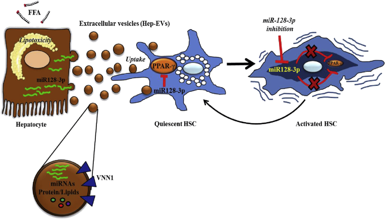
BACKGROUND&AIMS: Hepatic stellate cells (HSCs) play a key role in liver fibrosis in various chronic liver disorders including nonalcoholic fatty liver disease (NAFLD). The development of liver fibrosis requires a phenotypic switch from quiescent to activated HSCs. The triggers for HSCs activation in NAFLD remain poorly understood. We investigated the role and molecular mechanism of extracellular vesicles (EVs) released by hepatocytes during lipotoxicity in modulation of HSC phenotype.

METHODS

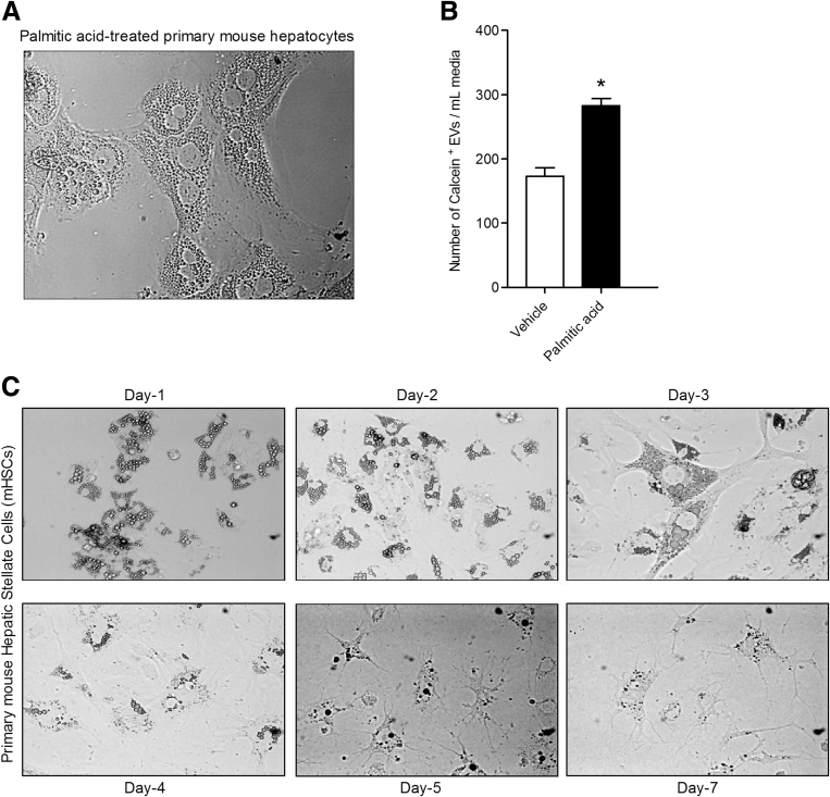
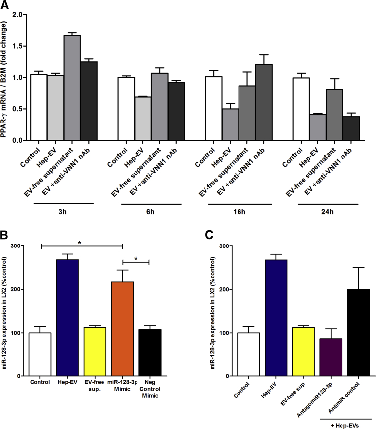
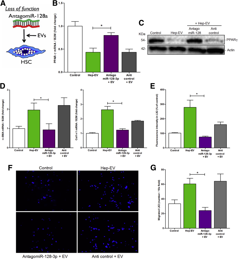
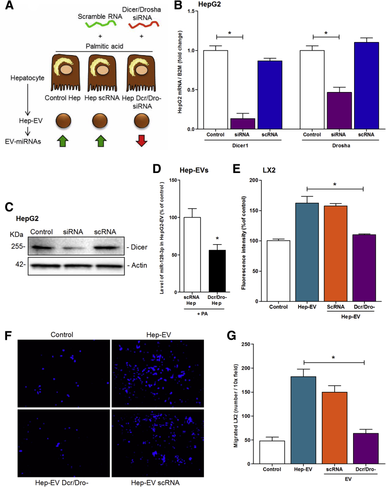
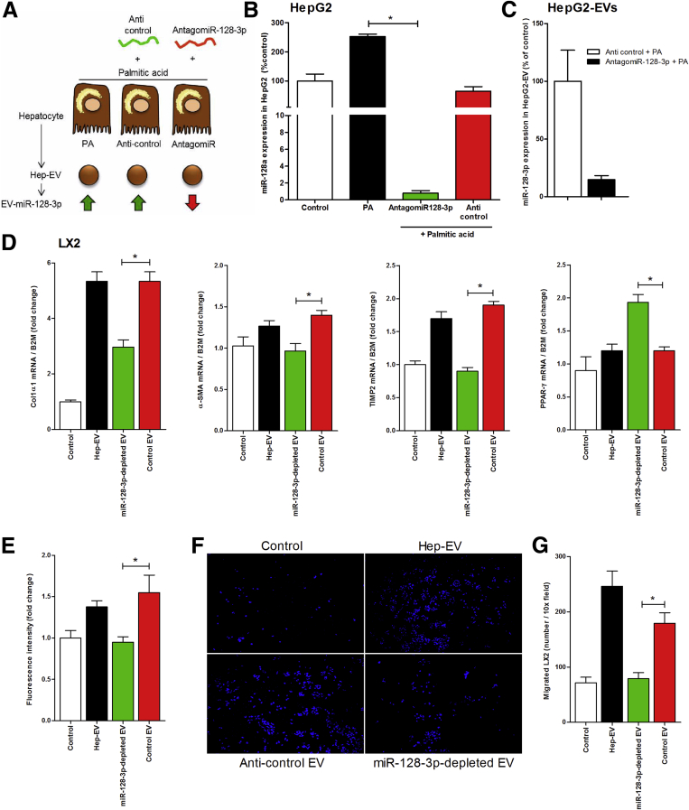
EVs were isolated from fat-laden hepatocytes by differential centrifugation and incubated with HSCs. EV internalization and HSCs activation, migration and proliferation were assessed. Loss- and gain-of-functions studies were performed to explore the potential role of PPAR-γ-targeting miRNAs carried by EVs into HSC.

RESULTS

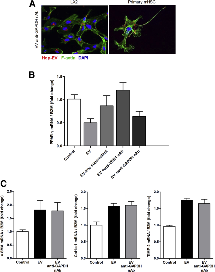
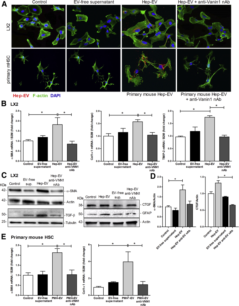
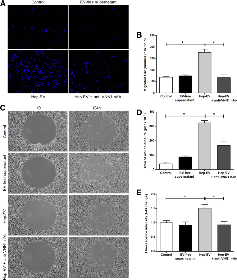
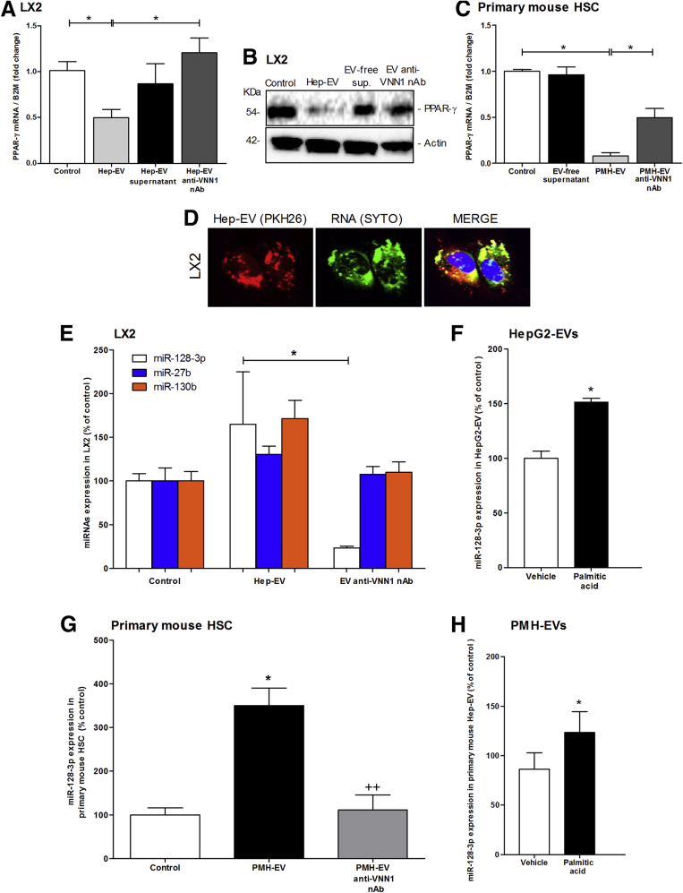
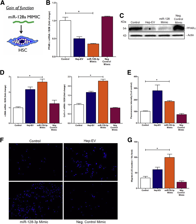
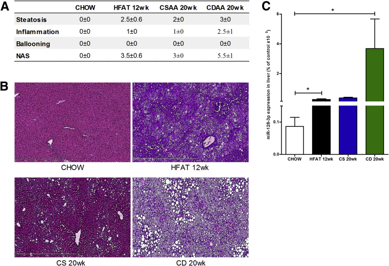
Hepatocyte-derived EVs released during lipotoxicity are efficiently internalized by HSCs resulting in their activation, as shown by marked up-regulation of pro-fibrogenic genes (Collagen-I, α-SMA and TIMP-2), proliferation, chemotaxis and wound healing responses. These changes were associated with miRNAs shuttled by EVs and suppression of PPAR-γ expression in HSC. Hepatocyte-derived EVs miRNA content included various miRNAs that are known inhibitors of PPAR-γ expression with miR-128-3p being the most effectively transferred. Furthermore loss- and gain-of-function studies identified miR-128-3p as a central modulator of the effects of EVs on PPAR-γ inhibition and HSC activation.

CONCLUSION

Our findings demonstrate a link between fat-laden hepatocyte-derived EVs and liver fibrosis and have potential implications for the development of novel anti-fibrotic targets for NAFLD and other fibrotic diseases.

摘要

背景与目的

肝星状细胞(HSCs)在包括非酒精性脂肪性肝病(NAFLD)在内的各种慢性肝脏疾病的肝纤维化过程中起关键作用。肝纤维化的发展需要肝星状细胞从静止状态转变为激活状态。NAFLD中肝星状细胞激活的触发因素仍知之甚少。我们研究了脂毒性期间肝细胞释放的细胞外囊泡(EVs)在调节肝星状细胞表型中的作用及分子机制。

方法

通过差速离心从富含脂肪的肝细胞中分离出细胞外囊泡,并与肝星状细胞一起孵育。评估细胞外囊泡的内化以及肝星状细胞的激活、迁移和增殖情况。进行功能缺失和功能获得研究,以探索细胞外囊泡携带的靶向过氧化物酶体增殖物激活受体γ(PPAR-γ)的微小RNA(miRNAs)在肝星状细胞中的潜在作用。

结果

脂毒性期间肝细胞衍生的细胞外囊泡被肝星状细胞有效内化,导致其激活,表现为促纤维化基因(I型胶原、α-平滑肌肌动蛋白和金属蛋白酶组织抑制因子-2)显著上调、增殖、趋化性和伤口愈合反应增强。这些变化与细胞外囊泡转运的微小RNA以及肝星状细胞中PPAR-γ表达的抑制有关。肝细胞衍生的细胞外囊泡微小RNA含量包括多种已知的PPAR-γ表达抑制剂微小RNA,其中miR-128-3p是最易转移的。此外,功能缺失和功能获得研究确定miR-128-3p是细胞外囊泡对PPAR-γ抑制和肝星状细胞激活作用的核心调节因子。

结论

我们的研究结果表明富含脂肪的肝细胞衍生的细胞外囊泡与肝纤维化之间存在联系,对NAFLD和其他纤维化疾病新型抗纤维化靶点的开发具有潜在意义。

https://cdn.ncbi.nlm.nih.gov/pmc/blobs/ea46/5301544/23e668bbaff1/gr9.jpg
https://cdn.ncbi.nlm.nih.gov/pmc/blobs/ea46/5301544/8fabd20c6b5f/fx1.jpg
https://cdn.ncbi.nlm.nih.gov/pmc/blobs/ea46/5301544/7c78a7dc3358/fx2.jpg
https://cdn.ncbi.nlm.nih.gov/pmc/blobs/ea46/5301544/436464882c98/fx3.jpg
https://cdn.ncbi.nlm.nih.gov/pmc/blobs/ea46/5301544/f428b67dee26/gr1.jpg
https://cdn.ncbi.nlm.nih.gov/pmc/blobs/ea46/5301544/892ea8e24d11/gr2.jpg
https://cdn.ncbi.nlm.nih.gov/pmc/blobs/ea46/5301544/3cc86ab5e7ad/gr3.jpg
https://cdn.ncbi.nlm.nih.gov/pmc/blobs/ea46/5301544/0108612c42c1/gr4.jpg
https://cdn.ncbi.nlm.nih.gov/pmc/blobs/ea46/5301544/b4f749a50034/gr5.jpg
https://cdn.ncbi.nlm.nih.gov/pmc/blobs/ea46/5301544/a89ee33bd8e0/gr6.jpg
https://cdn.ncbi.nlm.nih.gov/pmc/blobs/ea46/5301544/70fc4d804acc/gr7.jpg
https://cdn.ncbi.nlm.nih.gov/pmc/blobs/ea46/5301544/fd00cf0853aa/gr8.jpg
https://cdn.ncbi.nlm.nih.gov/pmc/blobs/ea46/5301544/23e668bbaff1/gr9.jpg

相似文献

1
Lipid-induced hepatocyte-derived extracellular vesicles regulate hepatic stellate cell via microRNAs targeting PPAR-γ.脂质诱导的肝细胞衍生细胞外囊泡通过靶向PPAR-γ的微小RNA调节肝星状细胞。
Cell Mol Gastroenterol Hepatol. 2015 Nov 1;1(6):646-663.e4. doi: 10.1016/j.jcmgh.2015.07.007.
2
Human induced pluripotent stem cell-derived extracellular vesicles reduce hepatic stellate cell activation and liver fibrosis.人诱导多能干细胞衍生的细胞外囊泡可减少肝星状细胞的激活和肝纤维化。
JCI Insight. 2019 Jun 11;5(14):125652. doi: 10.1172/jci.insight.125652.
3
Therapeutic effects of serum extracellular vesicles in liver fibrosis.血清细胞外囊泡在肝纤维化中的治疗作用。
J Extracell Vesicles. 2018 Apr 17;7(1):1461505. doi: 10.1080/20013078.2018.1461505. eCollection 2018.
4
Comprehensive characterization of hepatocyte-derived extracellular vesicles identifies direct miRNA-based regulation of hepatic stellate cells and DAMP-based hepatic macrophage IL-1β and IL-17 upregulation in alcoholic hepatitis mice.全面表征肝细胞衍生的细胞外囊泡,鉴定其对酒精性肝炎小鼠肝星状细胞的直接 miRNA 调控和基于 DAMPs 的肝巨噬细胞 IL-1β和 IL-17 的上调作用。
J Mol Med (Berl). 2020 Jul;98(7):1021-1034. doi: 10.1007/s00109-020-01926-7. Epub 2020 Jun 18.
5
Extracellular vesicles derived from fat-laden hepatocytes undergoing chemical hypoxia promote a pro-fibrotic phenotype in hepatic stellate cells.脂变肝细胞来源的细胞外囊泡在化学缺氧下促进肝星状细胞的促纤维化表型。
Biochim Biophys Acta Mol Basis Dis. 2020 Oct 1;1866(10):165857. doi: 10.1016/j.bbadis.2020.165857. Epub 2020 Jun 5.
6
MicroRNA-101 suppresses liver fibrosis by targeting the TGFβ signalling pathway.MicroRNA-101 通过靶向 TGFβ 信号通路抑制肝纤维化。
J Pathol. 2014 Sep;234(1):46-59. doi: 10.1002/path.4373. Epub 2014 Jun 17.
7
Hepatocyte-derived extracellular vesicles promote endothelial inflammation and atherogenesis via microRNA-1.肝细胞衍生的细胞外囊泡通过 microRNA-1 促进内皮炎症和动脉粥样硬化形成。
J Hepatol. 2020 Jan;72(1):156-166. doi: 10.1016/j.jhep.2019.09.014. Epub 2019 Sep 27.
8
Aflatoxin B-exposed hepatocyte-derived extracellular vesicles: Initiating hepatic stellate cell-mediated liver fibrosis through a p53-Parkin-dependent mitophagy pathway.黄曲霉毒素 B 暴露的肝细胞衍生细胞外囊泡:通过 p53-Parkin 依赖的线粒体自噬途径引发肝星状细胞介导的肝纤维化。
Ecotoxicol Environ Saf. 2024 Jun 1;277:116363. doi: 10.1016/j.ecoenv.2024.116363. Epub 2024 Apr 25.
9
Human Liver Stem Cell-Derived Extracellular Vesicles Target Hepatic Stellate Cells and Attenuate Their Pro-fibrotic Phenotype.人肝干细胞衍生的细胞外囊泡靶向肝星状细胞并减弱其促纤维化表型。
Front Cell Dev Biol. 2021 Nov 2;9:777462. doi: 10.3389/fcell.2021.777462. eCollection 2021.
10
Extracellular Vesicles from Steatotic Hepatocytes Provoke Pro-Fibrotic Responses in Cultured Stellate Cells.脂肪变性肝细胞来源的细胞外囊泡在体外诱导肝星状细胞产生促纤维化反应。
Biomolecules. 2022 May 13;12(5):698. doi: 10.3390/biom12050698.

引用本文的文献

1
Dynamic crosstalk between HSCs and liver microenvironment: multicellular interactions in the regulation of liver fibrosis.肝星状细胞与肝脏微环境之间的动态串扰:肝脏纤维化调控中的多细胞相互作用
Front Cell Dev Biol. 2025 Jul 21;13:1635763. doi: 10.3389/fcell.2025.1635763. eCollection 2025.
2
Communication initiated by hepatocytes: The driver of HSC activation and liver fibrosis.肝细胞引发的通讯:肝星状细胞激活和肝纤维化的驱动因素
Hepatol Commun. 2025 Jul 14;9(8). doi: 10.1097/HC9.0000000000000753. eCollection 2025 Aug 1.
3
Extracellular Vesicles as Biomarkers in Chronic Hepatobiliary Diseases: An Overview of Their Interplay.

本文引用的文献

1
Circulating extracellular vesicles with specific proteome and liver microRNAs are potential biomarkers for liver injury in experimental fatty liver disease.具有特定蛋白质组和肝脏微小RNA的循环细胞外囊泡是实验性脂肪性肝病中肝损伤的潜在生物标志物。
PLoS One. 2014 Dec 3;9(12):e113651. doi: 10.1371/journal.pone.0113651. eCollection 2014.
2
MicroRNA function in the profibrogenic interplay upon chronic liver disease.微小RNA在慢性肝病促纤维化相互作用中的作用
Int J Mol Sci. 2014 May 27;15(6):9360-71. doi: 10.3390/ijms15069360.
3
Activated macrophages release microvesicles containing polarized M1 or M2 mRNAs.
细胞外囊泡作为慢性肝胆疾病的生物标志物:它们相互作用的概述
Int J Mol Sci. 2025 Jun 30;26(13):6333. doi: 10.3390/ijms26136333.
4
The Role of Extracellular Vesicles in the Pathogenesis of Metabolic Dysfunction-Associated Steatotic Liver Disease and Other Liver Diseases.细胞外囊泡在代谢功能障碍相关脂肪性肝病及其他肝脏疾病发病机制中的作用
Int J Mol Sci. 2025 May 23;26(11):5033. doi: 10.3390/ijms26115033.
5
BMDM-derived ORP8 suppresses lipotoxicity and inflammation by relieving endoplasmic reticulum stress in mice with MASH.骨髓来源的ORP8通过减轻MASH小鼠的内质网应激来抑制脂毒性和炎症。
Mol Med. 2025 May 30;31(1):213. doi: 10.1186/s10020-025-01275-6.
6
Neutrophil-lymphocyte ratio is associated with worse outcomes in patients with cirrhosis: insights from the PRO-LIVER Registry.中性粒细胞与淋巴细胞比值与肝硬化患者的不良预后相关:来自PRO-LIVER注册研究的见解。
Intern Emerg Med. 2025 May 20. doi: 10.1007/s11739-025-03955-x.
7
Extracellular Vesicle-Mediated Network in the Pathogenesis of Obesity, Diabetes, Steatotic Liver Disease, and Cardiovascular Disease.细胞外囊泡介导的网络在肥胖症、糖尿病、脂肪性肝病和心血管疾病发病机制中的作用
Diabetes Metab J. 2025 May;49(3):348-367. doi: 10.4093/dmj.2025.0184. Epub 2025 May 1.
8
Characterization of Extracellular Vesicles Derived From Human Precision-Cut Liver Slices in Metabolic Dysfunction-Associated Steatotic Liver Disease.代谢功能障碍相关脂肪性肝病中源自人精密肝切片的细胞外囊泡的表征
J Extracell Biol. 2025 Apr 30;4(5):e70043. doi: 10.1002/jex2.70043. eCollection 2025 May.
9
Roles of extracellular vesicles from different origins in metabolic-associated fatty liver disease: progress and perspectives.不同来源的细胞外囊泡在代谢相关脂肪性肝病中的作用:进展与展望
Front Immunol. 2025 Mar 10;16:1544012. doi: 10.3389/fimmu.2025.1544012. eCollection 2025.
10
Therapeutic Actions of Hepatocyte Extracellular Vesicles in a Murine Model of Diet-Induced Steatohepatitis with Fibrosis.肝细胞外囊泡在饮食诱导的伴有纤维化的脂肪性肝炎小鼠模型中的治疗作用
Biomedicines. 2025 Jan 23;13(2):274. doi: 10.3390/biomedicines13020274.
活化的巨噬细胞释放含有极化的 M1 或 M2 mRNA 的微泡。
J Leukoc Biol. 2014 May;95(5):817-825. doi: 10.1189/jlb.0913485. Epub 2013 Dec 30.
4
Microvesicle-shuttled miR-130b reduces fat deposition in recipient primary cultured porcine adipocytes by inhibiting PPAR-g expression.微泡转运的 miR-130b 通过抑制 PPAR-γ 表达减少受体原代培养猪脂肪细胞中的脂肪沉积。
J Cell Physiol. 2014 May;229(5):631-9. doi: 10.1002/jcp.24486.
5
Dynamic expression of miR-126* and its effects on proliferation and contraction of hepatic stellate cells.miR-126* 的动态表达及其对肝星状细胞增殖和收缩的影响。
FEBS Lett. 2013 Nov 29;587(23):3792-801. doi: 10.1016/j.febslet.2013.09.047. Epub 2013 Oct 16.
6
Lipid-induced toxicity stimulates hepatocytes to release angiogenic microparticles that require Vanin-1 for uptake by endothelial cells.脂质诱导的毒性刺激肝细胞释放血管生成的微颗粒,这些微颗粒需要 Vanin-1 才能被内皮细胞摄取。
Sci Signal. 2013 Oct 8;6(296):ra88. doi: 10.1126/scisignal.2004512.
7
Amplified inhibition of stellate cell activation pathways by PPAR-γ, RAR and RXR agonists.PPAR-γ、RAR 和 RXR 激动剂对星状细胞激活途径的放大抑制作用。
PLoS One. 2013 Oct 1;8(10):e76541. doi: 10.1371/journal.pone.0076541. eCollection 2013.
8
Endothelial microparticle-mediated transfer of MicroRNA-126 promotes vascular endothelial cell repair via SPRED1 and is abrogated in glucose-damaged endothelial microparticles.内皮细胞微粒介导的 MicroRNA-126 转移通过 SPRED1 促进血管内皮细胞修复,并且在葡萄糖损伤的内皮细胞微粒中被阻断。
Circulation. 2013 Oct 29;128(18):2026-38. doi: 10.1161/CIRCULATIONAHA.113.001720. Epub 2013 Sep 6.
9
Microvesicle-mediated transfer of microRNA-150 from monocytes to endothelial cells promotes angiogenesis.微小囊泡介导的微小 RNA-150 从单核细胞向内皮细胞的转移促进血管生成。
J Biol Chem. 2013 Aug 9;288(32):23586-96. doi: 10.1074/jbc.M113.489302. Epub 2013 Jun 13.
10
Extracellular vesicles: exosomes, microvesicles, and friends.细胞外囊泡:外泌体、微囊泡及其他。
J Cell Biol. 2013 Feb 18;200(4):373-83. doi: 10.1083/jcb.201211138.