Higashide Tomomi, Ohkubo Shinji, Hangai Masanori, Ito Yasuki, Shimada Noriaki, Ohno-Matsui Kyoko, Terasaki Hiroko, Sugiyama Kazuhisa, Chew Paul, Li Kenneth K W, Yoshimura Nagahisa
Department of Ophthalmology and Visual Science, Kanazawa University Graduate School of Medical Science, Kanazawa, Japan.
Department of Ophthalmology, Saitama Medical University, Saitama, Japan.
PLoS One. 2016 Jan 27;11(1):e0147782. doi: 10.1371/journal.pone.0147782. eCollection 2016.
To identify the factors which significantly contribute to the thickness variabilities in macular retinal layers measured by optical coherence tomography with or without magnification correction of analytical areas in normal subjects.
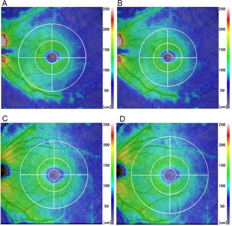
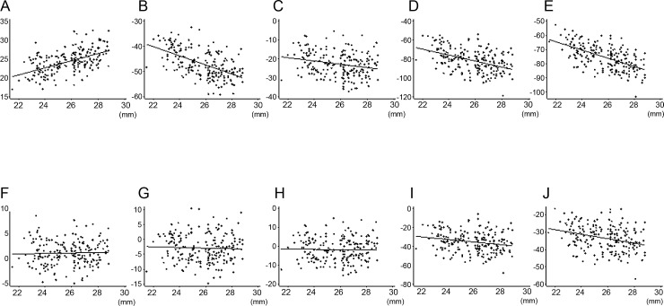
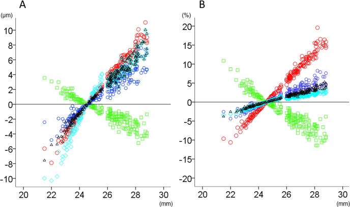
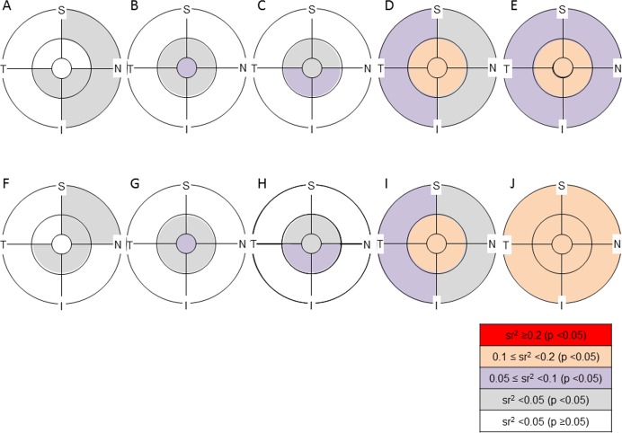
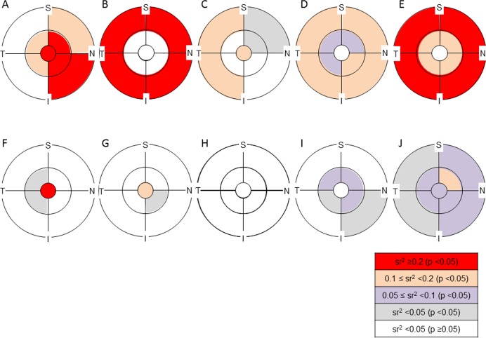
The thickness of retinal layers {retinal nerve fiber layer (RNFL), ganglion cell layer plus inner plexiform layer (GCLIPL), RNFL plus GCLIPL (ganglion cell complex, GCC), total retina, total retina minus GCC (outer retina)} were measured by macular scans (RS-3000, NIDEK) in 202 eyes of 202 normal Asian subjects aged 20 to 60 years. The analytical areas were defined by three concentric circles (1-, 3- and 6-mm nominal diameters) with or without magnification correction. For each layer thickness, a semipartial correlation (sr) was calculated for explanatory variables including age, gender, axial length, corneal curvature, and signal strength index.
Outer retinal thickness was significantly thinner in females than in males (sr2, 0.07 to 0.13) regardless of analytical areas or magnification correction. Without magnification correction, axial length had a significant positive sr with RNFL (sr2, 0.12 to 0.33) and a negative sr with GCLIPL (sr2, 0.22 to 0.31), GCC (sr2, 0.03 to 0.17), total retina (sr2, 0.07 to 0.17) and outer retina (sr2, 0.16 to 0.29) in multiple analytical areas. The significant sr in RNFL, GCLIPL and GCC became mostly insignificant following magnification correction.
The strong correlation between the thickness of inner retinal layers and axial length appeared to result from magnification effects. Outer retinal thickness may differ by gender and axial length independently of magnification correction.
确定在正常受试者中,通过光学相干断层扫描测量黄斑视网膜各层厚度时,有无分析区域放大校正情况下,对厚度变异性有显著影响的因素。
对202名年龄在20至60岁的正常亚洲受试者的202只眼睛进行黄斑扫描(RS - 3000,尼德克),测量视网膜各层(视网膜神经纤维层(RNFL)、神经节细胞层加内丛状层(GCLIPL)、RNFL加GCLIPL(神经节细胞复合体,GCC)、整个视网膜、整个视网膜减去GCC(外层视网膜))的厚度。分析区域由三个同心圆(标称直径分别为1、3和6毫米)定义,有或没有放大校正。对于每层厚度,计算与包括年龄、性别、眼轴长度、角膜曲率和信号强度指数在内的解释变量的半偏相关(sr)。
无论分析区域或放大校正情况如何,女性的外层视网膜厚度均显著薄于男性(sr²,0.07至0.13)。在没有放大校正的情况下,眼轴长度与多个分析区域的RNFL呈显著正相关(sr²,0.12至0.33),与GCLIPL呈负相关(sr²,0.22至0.31),与GCC呈负相关(sr²,0.03至0.17),与整个视网膜呈负相关(sr²,0.07至0.17),与外层视网膜呈负相关(sr²,0.16至0.29)。放大校正后,RNFL、GCLIPL和GCC中的显著sr大多不再显著。
视网膜内层厚度与眼轴长度之间的强相关性似乎是由放大效应导致的。外层视网膜厚度可能因性别和眼轴长度不同,且与放大校正无关。