Eppenberger Leila Sara, Li Chi, Wong Damon, Tan Bingyao, Garhöfer Gerhard, Hilal Saima, Chong Eddie, Toh An Qi, Venketasubramanian Narayanaswamy, Chen Christopher Li-Hsian, Schmetterer Leopold, Chua Jacqueline
Singapore Eye Research Institute, Singapore National Eye Centre, Singapore, Singapore.
SERI-NTU Advanced Ocular Engineering (STANCE), Singapore, Singapore.
Alzheimers Res Ther. 2024 Dec 23;16(1):273. doi: 10.1186/s13195-024-01627-0.
Dementia poses a significant burden on healthcare systems. Early identification of individuals at risk for cognitive decline is crucial. The retina, an extension of the central nervous system, reflects neurodegenerative changes. Optical coherence tomography (OCT) is a non-invasive tool for assessing retinal health and has shown promise in predicting cognitive decline. However, prior studies produced mixed results.
This study investigated a large cohort (n = 490) of Asian individuals attending memory clinics. Participants underwent comprehensive neuropsychological testing annually for five years. Retinal thickness was measured by OCT at baseline. We assessed the association between baseline retinal thickness and subsequent cognitive decline.
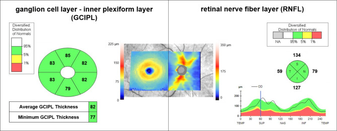
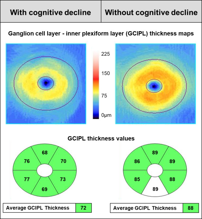
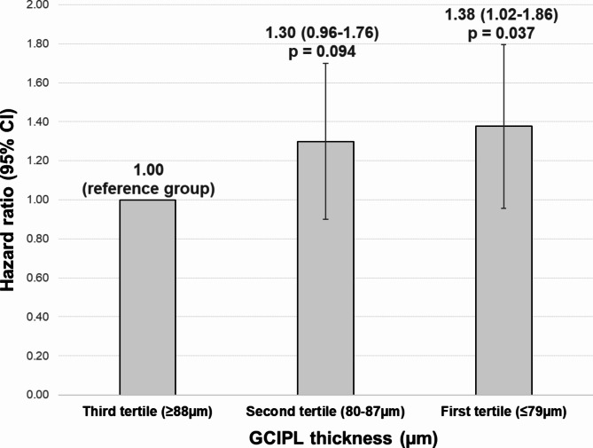
Participants with a significantly thinner macular ganglion cell-inner plexiform layer (GCIPL) at baseline (≤ 79 μm) had a 38% greater risk of cognitive decline compared to those who did not (≥ 88 μm; p = 0.037). In a multivariable model accounting for age, education, cerebrovascular disease status, hypertension, hyperlipidemia, diabetes and smoking, thinner GCIPL was associated with an increased risk of cognitive decline (hazard ratio = 1.14, 95% CI = 1.01-1.30, p = 0.035). Retinal nerve fiber layer (RNFL) thickness was not associated with cognitive decline.
This study suggests that OCT-derived macular GCIPL thickness may be a valuable biomarker for identifying individuals at risk of cognitive decline. Our findings highlight GCIPL as a potentially more sensitive marker compared to RNFL thickness for detecting early neurodegenerative changes.
National Healthcare Group Domain-Specific Review Board (NHG DSRB) reference numbers DSRB Ref: 2018/01368. Name of the trial: Harmonisation project.
痴呆给医疗系统带来了沉重负担。尽早识别有认知能力下降风险的个体至关重要。视网膜作为中枢神经系统的延伸,反映了神经退行性变化。光学相干断层扫描(OCT)是一种评估视网膜健康的非侵入性工具,在预测认知能力下降方面已显示出前景。然而,先前的研究结果不一。
本研究调查了一大群(n = 490)前往记忆诊所就诊的亚洲个体。参与者在五年内每年接受全面的神经心理学测试。在基线时通过OCT测量视网膜厚度。我们评估了基线视网膜厚度与随后认知能力下降之间的关联。
基线时黄斑神经节细胞 - 内丛状层(GCIPL)明显更薄(≤ 79μm)的参与者与未出现这种情况(≥ 88μm)的参与者相比,认知能力下降的风险高38%(p = 0.037)。在一个考虑了年龄、教育程度、脑血管疾病状况、高血压、高脂血症、糖尿病和吸烟因素的多变量模型中,较薄的GCIPL与认知能力下降风险增加相关(风险比 = 1.14,95%置信区间 = 1.01 - 1.30,p = 0.035)。视网膜神经纤维层(RNFL)厚度与认知能力下降无关。
本研究表明,OCT得出的黄斑GCIPL厚度可能是识别有认知能力下降风险个体的有价值生物标志物。我们的研究结果突出了GCIPL作为一种比RNFL厚度在检测早期神经退行性变化方面可能更敏感的标志物。
国家医疗集团特定领域审查委员会(NHG DSRB)参考编号DSRB Ref: 2018/01368。试验名称:协调项目。