Jester James V, Parfitt Geraint J, Brown Donald J
Gavin Herbert Eye Institute, University of California, Irvine, CA, USA.
BMC Ophthalmol. 2015 Dec 17;15 Suppl 1(Suppl 1):156. doi: 10.1186/s12886-015-0132-x.
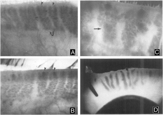
Meibomian gland dysfunction (MGD) is the major cause of evaporative dry eye disease (EDED) and dysfunction is widely thought to mechanistically involve ductal hyperkeratinization, plugging and obstruction. This review re-evaluates the role of hyperkeratinization in MGD based on more recent findings from mouse models. In these studies, eyelids from normal young and old mice or mice exposed to desiccating stress were evaluated by immunofluorescent tomography and 3-dimensional reconstruction to evaluate gland volume, expression of hyperkeratinization markers and cell proliferation or stimulated Raman scattering (SRS) microscopy to assess lipid quality. Results indicate that aging mice show dropout of meibomian glands with loss of gland volume and a forward migration of the mucocutaneous junction anterior to the gland orifice; similar age-related changes that are detected in human subjects. Atrophic glands also showed evidence of epithelial plugging of the orifice without the presence of hyperkeratinization. Mice exposed to desiccating stress showed hyperproliferation of the meibomian gland and ductal dilation suggesting a marked increase in lipid synthesis. Lipid quality was also affected in EDED mice with an increase in the protein content of lipid within the duct of the gland. Overall, age-related changes in the mouse show similar structural and functional correlates with that observed in clinical MGD without evidence of hyperkeratinization suggesting that gland atrophy may be a major cause of EDED. The response of the meibomian gland to desiccating stress also suggest that environmental conditions may accelerate or potentiate age-related changes.
睑板腺功能障碍(MGD)是蒸发型干眼疾病(EDED)的主要病因,人们普遍认为其功能障碍在机制上涉及导管过度角化、堵塞和梗阻。本综述基于小鼠模型的最新研究结果,重新评估了过度角化在MGD中的作用。在这些研究中,通过免疫荧光断层扫描和三维重建来评估正常年轻和老年小鼠或暴露于干燥应激的小鼠的眼睑,以评估腺体体积、过度角化标志物的表达和细胞增殖,或通过受激拉曼散射(SRS)显微镜来评估脂质质量。结果表明,衰老小鼠的睑板腺出现缺失,腺体体积减小,黏膜皮肤交界处向前迁移至腺体开口前方;在人类受试者中也检测到类似的年龄相关变化。萎缩的腺体也显示出开口处上皮堵塞的迹象,但没有过度角化。暴露于干燥应激的小鼠睑板腺出现过度增殖和导管扩张,提示脂质合成显著增加。EDED小鼠的脂质质量也受到影响,腺体导管内脂质的蛋白质含量增加。总体而言,小鼠的年龄相关变化与临床MGD中观察到的结构和功能相关性相似,没有过度角化的证据,这表明腺体萎缩可能是EDED的主要原因。睑板腺对干燥应激的反应也表明,环境条件可能加速或加剧年龄相关变化。