Höög Johanna L, Lacomble Sylvain, Bouchet-Marquis Cedric, Briggs Laura, Park Kristin, Hoenger Andreas, Gull Keith
Sir William Dunn School of Pathology, University of Oxford, Oxford, United Kingdom.
The Boulder Laboratory for 3D Electron Microscopy of Cells, Department of MCD Biology, University of Colorado, Boulder, Colorado, United States of America.
PLoS Negl Trop Dis. 2016 Jan 28;10(1):e0004312. doi: 10.1371/journal.pntd.0004312. eCollection 2016 Jan.
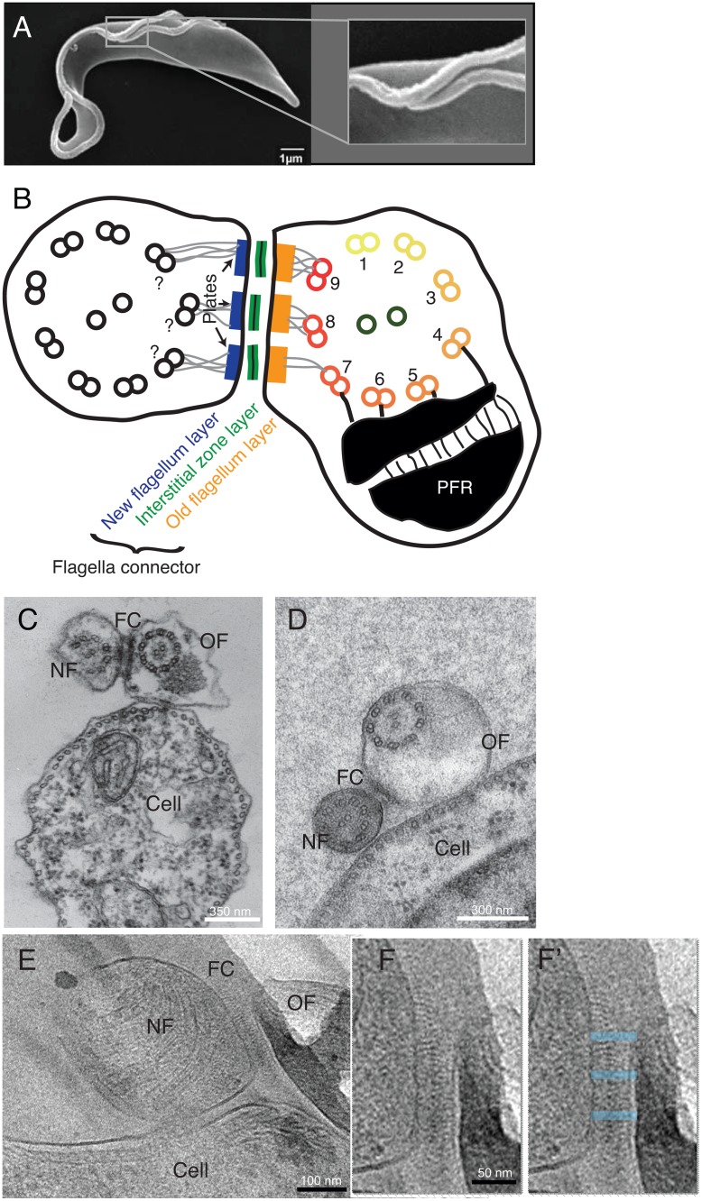
Cellular junctions are crucial for the formation of multicellular organisms, where they anchor cells to each other and/or supportive tissue and enable cell-to-cell communication. Some unicellular organisms, such as the parasitic protist Trypanosoma brucei, also have complex cellular junctions. The flagella connector (FC) is a three-layered transmembrane junction that moves with the growing tip of a new flagellum and attaches it to the side of the old flagellum. The FC moves via an unknown molecular mechanism, independent of new flagellum growth. Here we describe the detailed 3D architecture of the FC suggesting explanations for how it functions and its mechanism of motility.
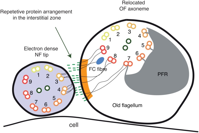
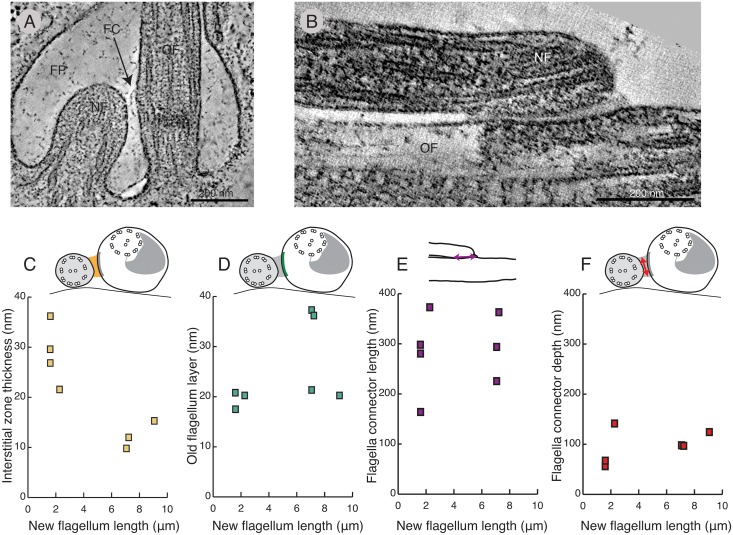
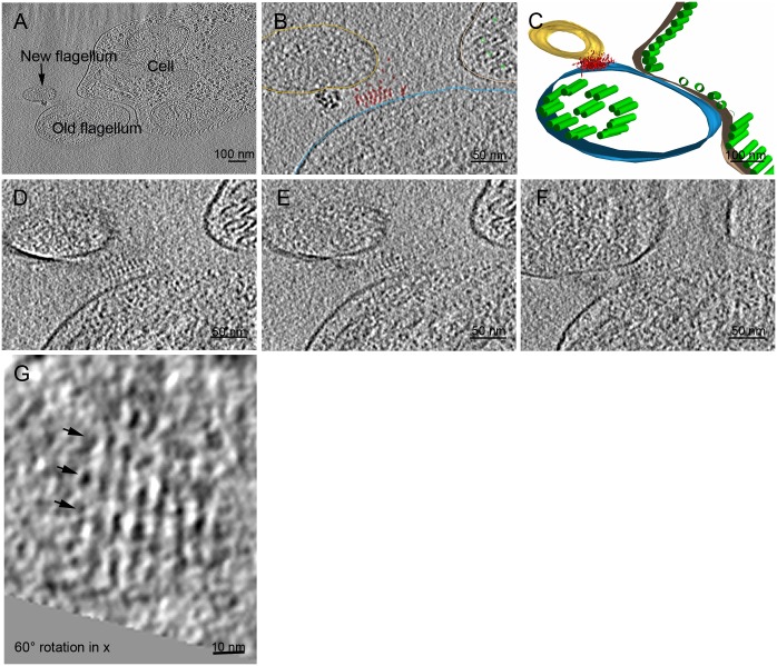
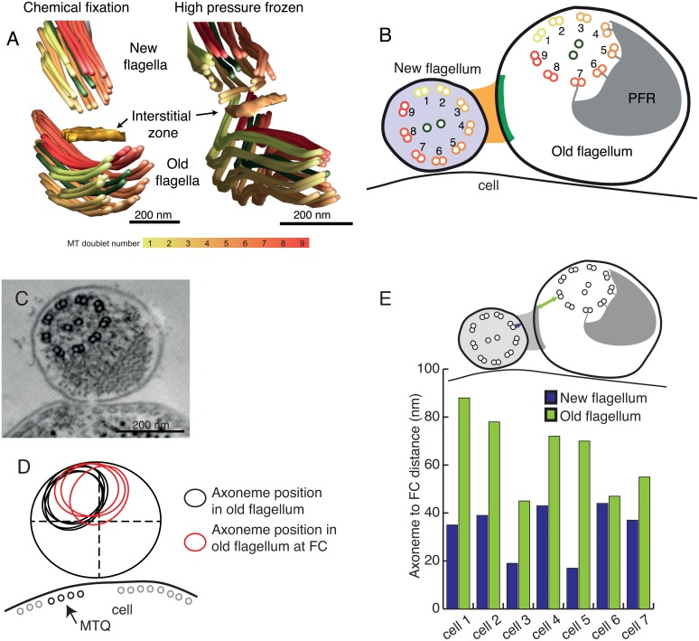
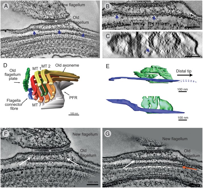
METHODOLOGY/PRINCIPAL FINDINGS: We have used a combination of electron tomography and cryo-electron tomography to reveal the 3D architecture of the FC. Cryo-electron tomography revealed layers of repetitive filamentous electron densities between the two flagella in the interstitial zone. Though the FC does not change in length and width during the growth of the new flagellum, the interstitial zone thickness decreases as the FC matures. This investigation also shows interactions between the FC layers and the axonemes of the new and old flagellum, sufficiently strong to displace the axoneme in the old flagellum. We describe a novel filament, the flagella connector fibre, found between the FC and the axoneme in the old flagellum.
CONCLUSIONS/SIGNIFICANCE: The FC is similar to other cellular junctions in that filamentous proteins bridge the extracellular space and are anchored to underlying cytoskeletal structures; however, it is built between different portions of the same cell and is unique because of its intrinsic motility. The detailed description of its structure will be an important tool to use in attributing structure / function relationships as its molecular components are discovered in the future. The FC is involved in the inheritance of cell shape, which is important for the life cycle of this human parasite.
细胞连接对于多细胞生物的形成至关重要,它将细胞彼此锚定以及/或者与支持组织相连,并实现细胞间通讯。一些单细胞生物,如寄生原生生物布氏锥虫,也具有复杂的细胞连接。鞭毛连接器(FC)是一种三层跨膜连接,它随着新鞭毛生长尖端移动,并将其附着于旧鞭毛的侧面。FC通过未知分子机制移动,独立于新鞭毛生长。在此,我们描述了FC的详细三维结构,对其功能方式及其运动机制提出了解释。
方法/主要发现:我们结合使用电子断层扫描和冷冻电子断层扫描来揭示FC的三维结构。冷冻电子断层扫描显示在间隙区域的两根鞭毛之间存在多层重复的丝状电子密度。尽管在新鞭毛生长过程中FC的长度和宽度不变,但随着FC成熟,间隙区域厚度减小。这项研究还显示了FC层与新旧鞭毛轴丝之间的相互作用,其强度足以使旧鞭毛中的轴丝移位。我们描述了一种在旧鞭毛的FC和轴丝之间发现的新型细丝,即鞭毛连接纤维。
结论/意义:FC与其他细胞连接类似,丝状蛋白桥接细胞外空间并锚定到潜在的细胞骨架结构;然而,它构建于同一细胞的不同部分之间,并且因其内在运动性而独特。随着其分子成分在未来被发现,对其结构的详细描述将成为确定结构/功能关系的重要工具。FC参与细胞形状的遗传,这对这种人体寄生虫的生命周期很重要。