Barrese James C, Aceros Juan, Donoghue John P
Department of Neurological Surgery, New Jersey Medical School, Rutgers University, Newark, NJ, USA. Department of Neuroscience and Brown Institute for Brain Science, Brown University, Providence, RI, USA.
J Neural Eng. 2016 Apr;13(2):026003. doi: 10.1088/1741-2560/13/2/026003. Epub 2016 Jan 29.
Signal attenuation is a major problem facing intracortical sensors for chronic neuroprosthetic applications. Many studies suggest that failure is due to gliosis around the electrode tips, however, mechanical and material causes of failure are often overlooked. The purpose of this study was to investigate the factors contributing to progressive signal decline by using scanning electron microscopy (SEM) to visualize structural changes in chronically implanted arrays and histology to examine the tissue response at corresponding implant sites.
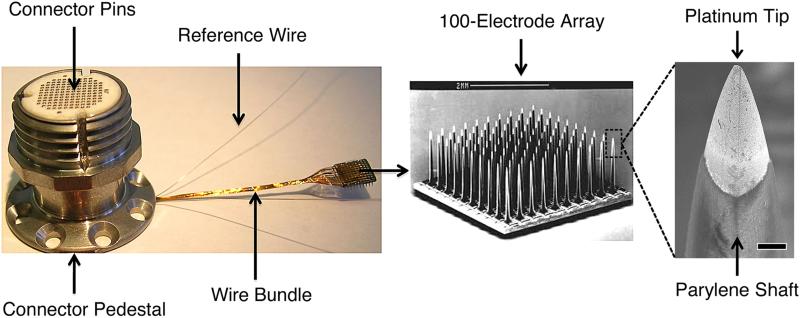
We examined eight chronically implanted intracortical microelectrode arrays (MEAs) explanted from non-human primates at times ranging from 37 to 1051 days post-implant. We used SEM, in vivo neural recordings, and histology (GFAP, Iba-1, NeuN). Three MEAs that were never implanted were also imaged as controls.
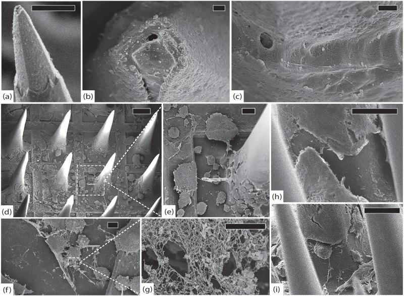
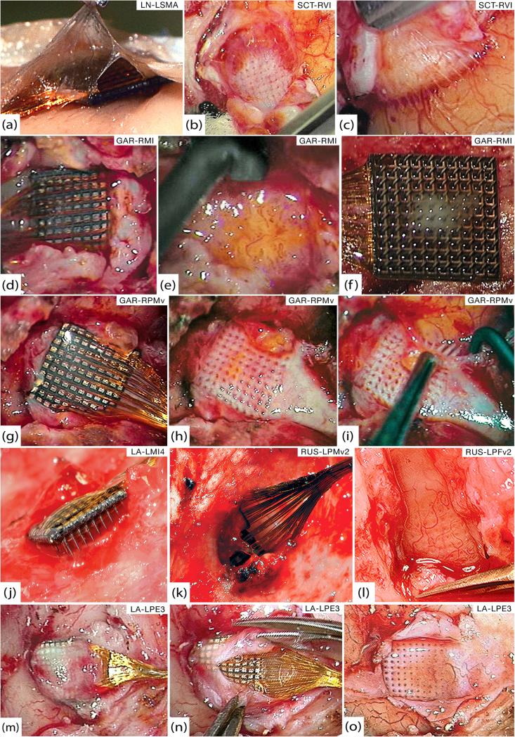
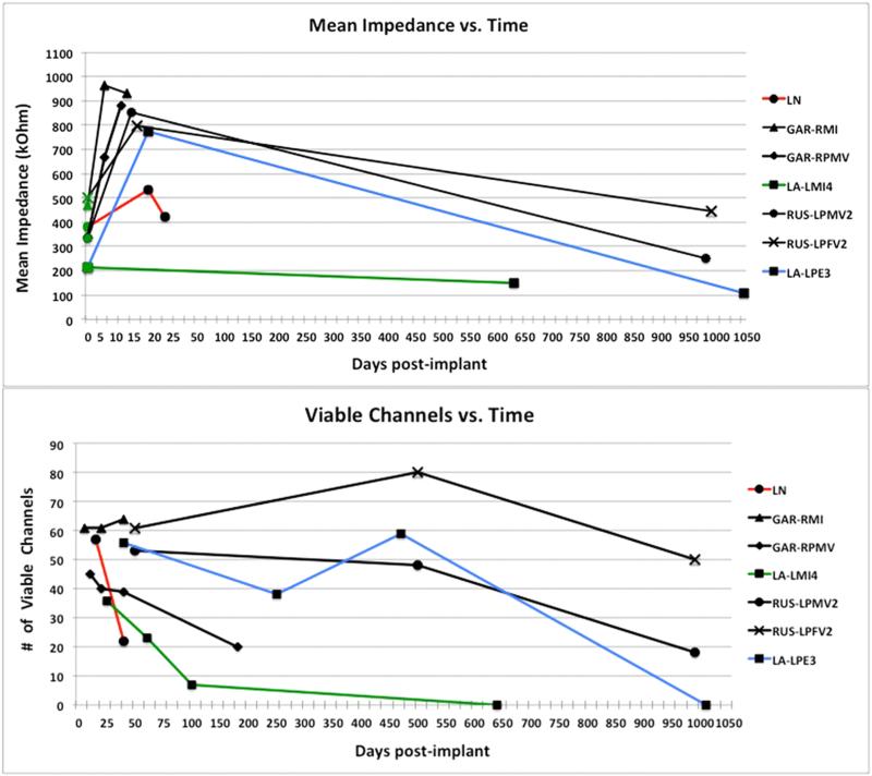
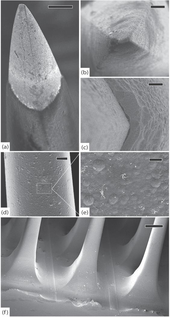
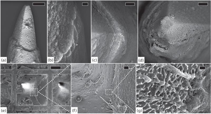
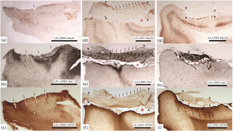
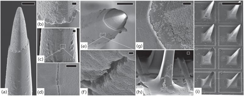
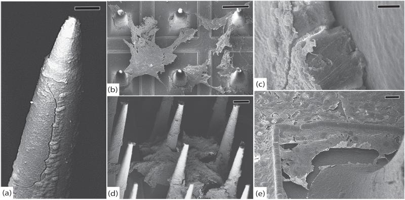
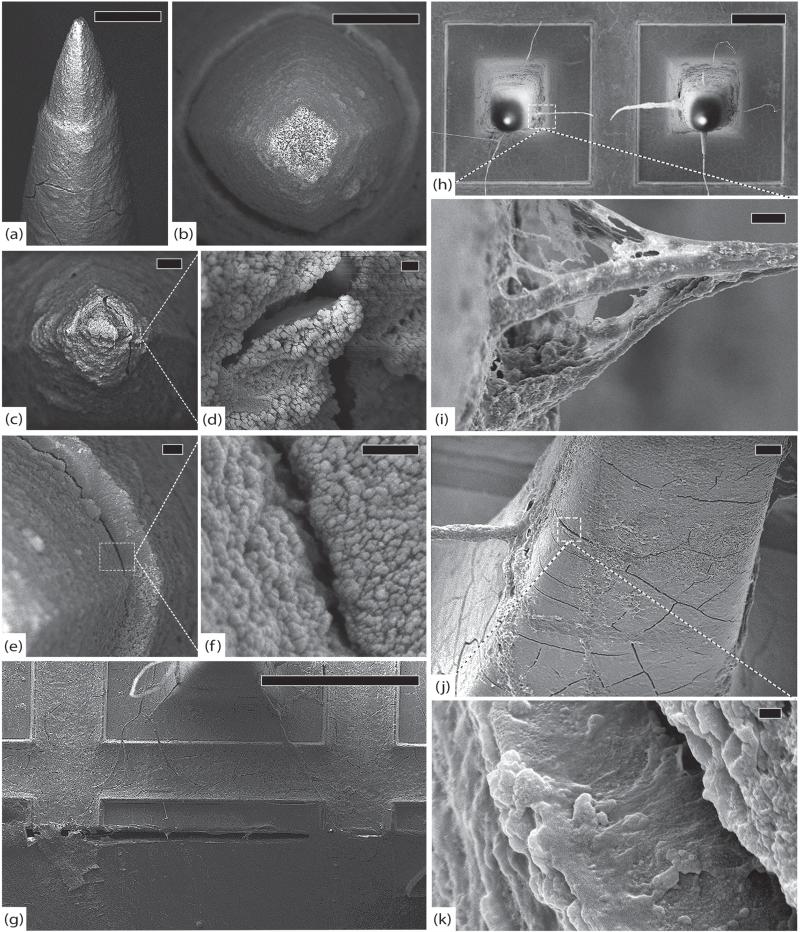
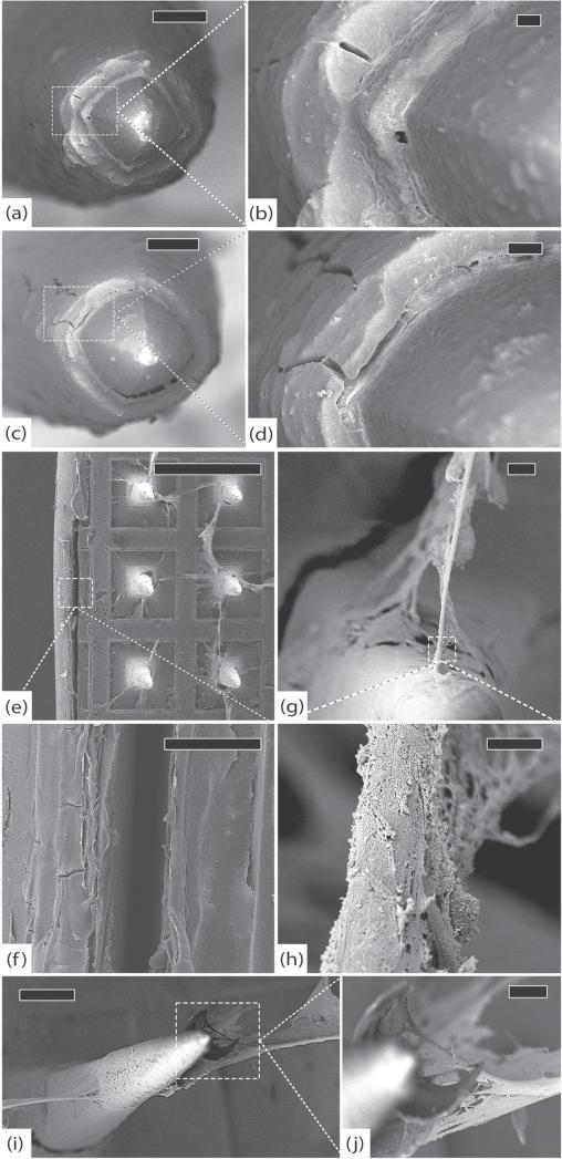
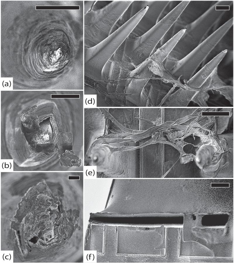
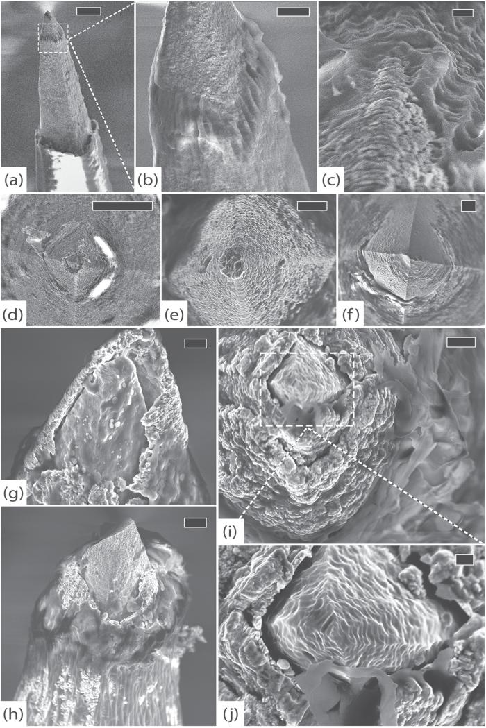
SEM revealed progressive corrosion of the platinum electrode tips and changes to the underlying silicon. The parylene insulation was prone to cracking and delamination, and in some instances the silicone elastomer also delaminated from the edges of the MEA. Substantial tissue encapsulation was observed and was often seen growing into defects in the platinum and parylene. These material defects became more common as the time in vivo increased. Histology at 37 and 1051 days post-implant showed gliosis, disruption of normal cortical architecture with minimal neuronal loss, and high Iba-1 reactivity, especially within the arachnoid and dura. Electrode tracts were either absent or barely visible in the cortex at 1051 days, but were seen in the fibrotic encapsulation material suggesting that the MEAs were lifted out of the brain. Neural recordings showed a progressive drop in impedance, signal amplitude, and viable channels over time.
These results provide evidence that signal loss in MEAs is truly multifactorial. Gliosis occurs in the first few months after implantation but does not prevent useful recordings for several years. Progressive meningeal fibrosis encapsulates and lifts MEAs out of the cortex while ongoing foreign body reactions lead to progressive degradation of the materials. Long-term impedance drops are due to the corrosion of platinum, cracking and delamination of parylene, and delamination of silicone elastomer. Oxygen radicals released by cells of the immune system likely mediate the degradation of these materials. Future MEA designs must address these problems through more durable insulation materials, more inert electrode alloys, and pharmacologic suppression of fibroblasts and leukocytes.
信号衰减是慢性神经假体应用中皮层内传感器面临的主要问题。许多研究表明,故障是由于电极尖端周围的胶质增生所致,然而,故障的机械和材料原因常常被忽视。本研究的目的是通过使用扫描电子显微镜(SEM)观察长期植入阵列的结构变化以及组织学检查相应植入部位的组织反应,来研究导致信号逐渐下降的因素。
我们检查了8个从非人灵长类动物体内取出的长期植入的皮层内微电极阵列(MEA),植入时间从37天到1051天不等。我们使用了SEM、体内神经记录和组织学(GFAP、Iba-1、NeuN)。还对3个从未植入的MEA进行了成像作为对照。
SEM显示铂电极尖端逐渐腐蚀以及其下方硅的变化。聚对二甲苯绝缘层容易出现裂纹和分层,在某些情况下,硅橡胶也会从MEA边缘分层。观察到大量组织包封,并且经常可见其生长到铂和聚对二甲苯的缺陷中。随着体内植入时间的增加,这些材料缺陷变得更加常见。植入后37天和1051天的组织学显示胶质增生、正常皮质结构破坏且神经元损失最小,以及高Iba-1反应性,尤其是在蛛网膜和硬脑膜内。在1051天时,电极轨迹在皮层中要么不存在要么几乎不可见,但在纤维化包封材料中可见,这表明MEA被从大脑中抬起。神经记录显示随着时间的推移,阻抗、信号幅度和可用通道逐渐下降。
这些结果提供了证据,表明MEA中的信号损失确实是多因素的。胶质增生在植入后的头几个月发生,但并不妨碍数年的有效记录。进行性脑膜纤维化将MEA包封并从皮层中抬起,同时持续的异物反应导致材料逐渐降解。长期阻抗下降是由于铂的腐蚀、聚对二甲苯的裂纹和分层以及硅橡胶的分层。免疫系统细胞释放的氧自由基可能介导了这些材料的降解。未来MEA的设计必须通过更耐用的绝缘材料、更惰性的电极合金以及对成纤维细胞和白细胞的药物抑制来解决这些问题。