Tartaglione T, Botto A, Sciandra M, Gaudino S, Danieli L, Parrilla C, Paludetti G, Colosimo C
Department of Radiological Sciences, Università Cattolica del Sacro Cuore, "A. Gemelli" Hospital, Roma, Italy.
Department of Head and Neck Surgery Otorhinolaryngology, Università Cattolica del Sacro Cuore, "A. Gemelli "Hospital, Roma, Italy.
Acta Otorhinolaryngol Ital. 2015 Oct;35(5):314-20. doi: 10.14639/0392-100X-693.
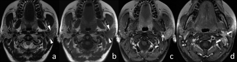
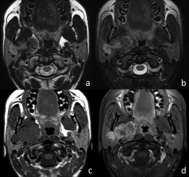
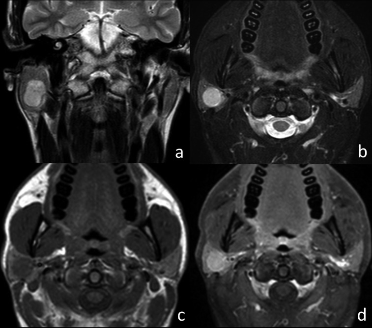
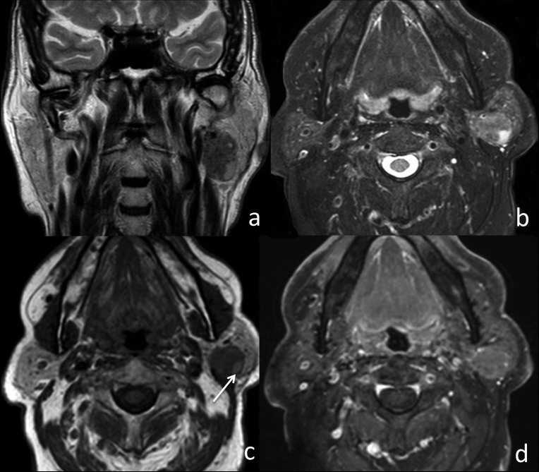
Our aim was to define typical magnetic resonance (MRI) findings in malignant and benign parotid tumours. This study is based on retrospective evaluation of pre-surgical MRI of 94 patients with parotid gland tumours. Histology results were available for all tumours. There were 69 cases of benign (73%) and 25 cases of malignant (27%) tumours, including 44 pleomorphic adenomas, 18 Warthin's tumours, 7 various benign tumours, 6 squamous cell carcinomas, 3 carcinoma ex pleomorphic adenomas, 2 mucoepidermoid carcinomas, 1 adenoid cystic carcinoma and 13 various malignant tumours. The following MRI parameters were evaluated: shape, site, size, margins, signal intensity (SI) on T1w and T2w images, contrast enhancement, signal of cystic content, presence or absence of a capsule, perineural spread, extraglandular growth pattern and cervical adenopathy. Statistical analysis was performed to identify the MRI findings most suggestive of malignancy, and to define the most typical MRI pattern of the most common histologies. Ill-defined margins (p < 0.001), adenopathies (p < 0.001) and infiltrative grown pattern (p < 0.001) were significantly predictive of malignancy. Typical findings of pleomorphic adenoma included hyperintensity on T2w images (p = 0.02), strong contrast enhancement (p < 0.001) and lobulated shape (p = 0.04). Typical findings of Warthin's tumour included hyperintense components on T1w images (p < 0.001), location in the parotid inferior process (p < 0.001) and mild or incomplete contrast enhancement (p = 0.01). SI on T1w and T2w images and contrast enhancement enables differential diagnosis between pleomorphic adenoma and Warthin's tumour.
我们的目的是明确腮腺良恶性肿瘤的典型磁共振成像(MRI)表现。本研究基于对94例腮腺肿瘤患者术前MRI的回顾性评估。所有肿瘤均有组织学结果。其中良性肿瘤69例(73%),恶性肿瘤25例(27%),包括44例多形性腺瘤、18例沃辛瘤、7例其他良性肿瘤、6例鳞状细胞癌、3例多形性腺瘤恶变、2例黏液表皮样癌、1例腺样囊性癌以及13例其他恶性肿瘤。评估了以下MRI参数:形态、位置、大小、边界、T1加权像和T2加权像上的信号强度(SI)、对比增强、囊内容物信号、有无包膜、神经周围扩散、腺外生长方式以及颈部淋巴结肿大。进行了统计学分析,以确定最提示恶性的MRI表现,并明确最常见组织学类型的最典型MRI模式。边界不清(p < 0.001)、淋巴结肿大(p < 0.001)和浸润性生长方式(p < 0.001)是恶性的显著预测指标。多形性腺瘤的典型表现包括T2加权像上高信号(p = 0.02)、强烈对比增强(p < 0.001)和分叶状形态(p = 0.04)。沃辛瘤的典型表现包括T1加权像上高信号成分(p < 0.001)、位于腮腺下极(p < 0.001)以及轻度或不完全对比增强(p = 0.01)。T1加权像和T2加权像上的SI以及对比增强有助于多形性腺瘤和沃辛瘤的鉴别诊断。