Singh Vedpal, Elamvazuthi Irraivan, Jeoti Varun, George John, Swain Akshya, Kumar Dileep
Centre for Intelligent Signal and Imaging Research (CISIR), Department of Electrical and Electronic Engineering, Universiti Teknologi PETRONAS, Bandar Seri Iskandar, 32610, Perak Darul Ridzuan, Malaysia.
Research Imaging Centre, University of Malaya, Kuala Lumpur, 50603, Malaysia.
Biomed Eng Online. 2016 Feb 2;15:13. doi: 10.1186/s12938-016-0129-6.
Anterior talofibular ligament (ATFL) is considered as the weakest ankle ligament that is most prone to injuries. Ultrasound imaging with its portable, non-invasive and non-ionizing radiation nature is increasingly being used for ATFL diagnosis. However, diagnosis of ATFL injuries requires its segmentation from ultrasound images that is a challenging task due to the existence of homogeneous intensity regions, homogeneous textures and low contrast regions in ultrasound images. To address these issues, this research has developed an efficient ATFL segmentation framework that would contribute to accurate and efficient diagnosis of ATFL injuries for clinical evaluation.
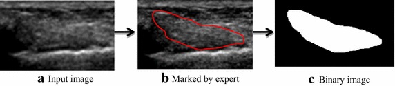
The developed framework comprises of five computational steps to segment the ATFL ligament region. Initially, region of interest is selected from the original image, which is followed by the adaptive histogram equalization to enhance the contrast level of the ultrasound image. The enhanced contrast image is further optimized by the particle swarm optimization algorithm. Thereafter, the optimized image is processed by the Chan-Vese method to extract the ATFL region through curve evolution; then the resultant image smoothed by morphological operation. The algorithm is tested on 25 subjects' datasets and the corresponding performance metrics are evaluated to demonstrate its clinical applicability.
The performance of the developed framework is evaluated based on various measurement metrics. It was found that estimated computational performance of the developed framework is 12 times faster than existing Chan-Vese method. Furthermore, the developed framework yielded the average sensitivity of 98.3 %, specificity of 96.6 % and accuracy of 96.8 % as compared to the manual segmentation. In addition, the obtained distance using Hausdorff is 14.2 pixels and similarity index by Jaccard is 91 %, which are indicating the enhanced performance whilst segmented area of ATFL region obtained from five normal (average Pixels-16,345.09), five tear (average Pixels-14,940.96) and five thickened (average Pixels-12,179.20) subjects' datasets show good performance of developed framework to be used in clinical practices.
On the basis of obtained results, the developed framework is computationally more efficient and more accurate with lowest rate of coefficient of variation (less than 5 %) that indicates the highest clinical significance of this research in the assessment of ATFL injuries.
距腓前韧带(ATFL)被认为是踝关节最薄弱且最易受伤的韧带。超声成像具有便携、无创和无电离辐射的特点,越来越多地用于ATFL损伤的诊断。然而,ATFL损伤的诊断需要从超声图像中分割出该韧带,由于超声图像中存在强度均匀区域、纹理均匀区域和低对比度区域,这是一项具有挑战性的任务。为了解决这些问题,本研究开发了一种高效的ATFL分割框架,有助于对ATFL损伤进行准确、高效的诊断,以用于临床评估。
所开发的框架包括五个计算步骤来分割ATFL韧带区域。首先,从原始图像中选择感兴趣区域,然后进行自适应直方图均衡化以提高超声图像的对比度。通过粒子群优化算法对增强后的对比度图像进一步优化。此后,利用Chan-Vese方法对优化后的图像进行处理,通过曲线演化提取ATFL区域;然后对所得图像进行形态学操作平滑处理。该算法在25名受试者的数据集上进行测试,并评估相应的性能指标以证明其临床适用性。
基于各种测量指标对所开发框架的性能进行评估。结果发现,所开发框架的估计计算性能比现有的Chan-Vese方法快12倍。此外,与手动分割相比,所开发框架的平均灵敏度为98.3%,特异性为96.6%,准确率为96.8%。此外,使用豪斯多夫距离得到的结果为14.2像素,杰卡德相似指数为91%,这表明在分割区域性能有所提高,同时从五名正常(平均像素数-16345.09)、五名撕裂(平均像素数-14940.96)和五名增厚(平均像素数-12179.20)受试者的数据集中获得的ATFL区域显示出所开发框架在临床实践中的良好性能。
基于所得结果,所开发的框架在计算上更高效、更准确,变异系数最低(小于5%),这表明本研究在ATFL损伤评估中具有最高的临床意义。