Swiecicki Jean-Marie, Thiebaut Frédéric, Di Pisa Margherita, Gourdin-Bertin Simon, Tailhades Julien, Mansuy Christelle, Burlina Fabienne, Chwetzoff Serge, Trugnan Germain, Chassaing Gérard, Lavielle Solange
Sorbonne Universités, UPMC Univ Paris 06, LBM, 4, Place Jussieu, 75005 Paris, France.
Ecole Normale Supérieure - PSL research University, Département de Chimie, 24 Rue Lhomond, 75005 Paris, France.
Sci Rep. 2016 Feb 3;6:20237. doi: 10.1038/srep20237.
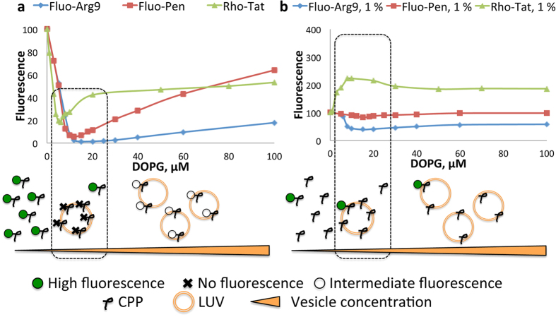
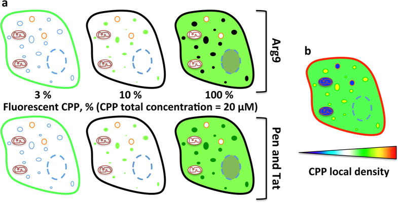
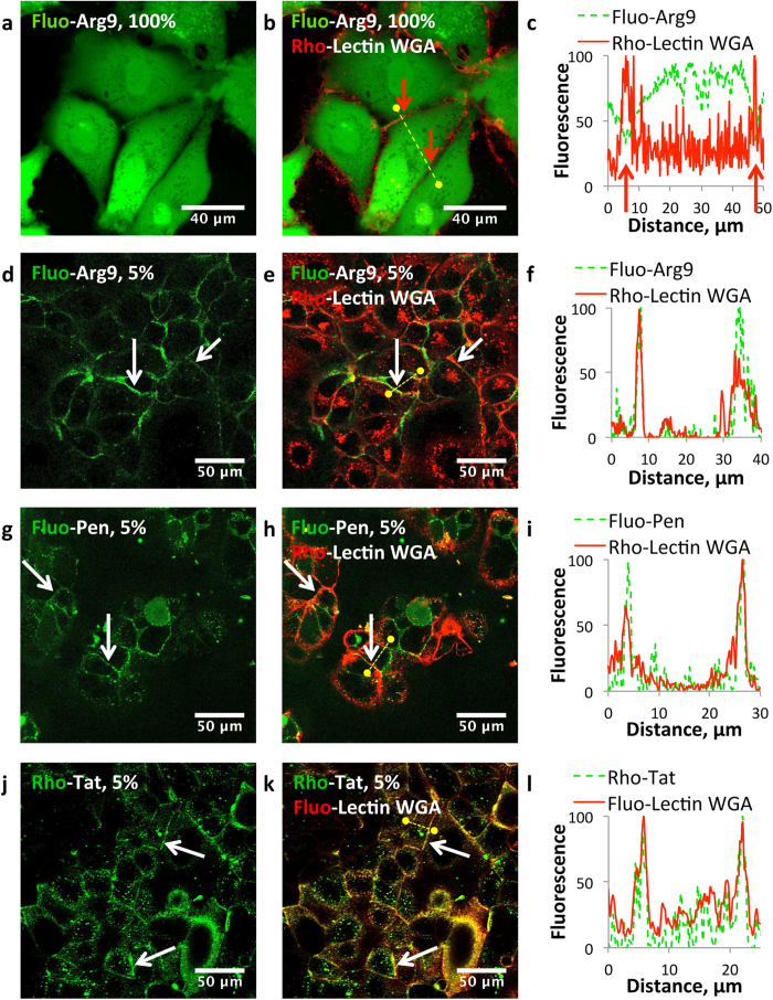
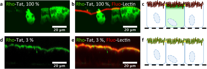
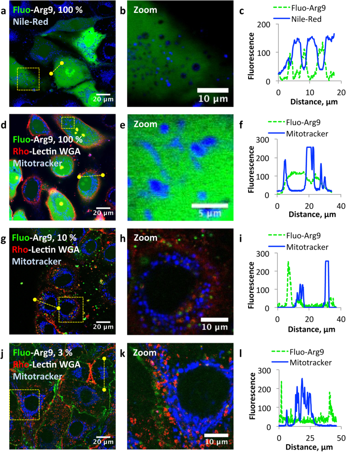
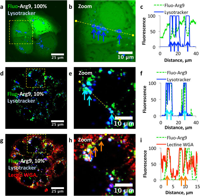
Confocal laser scanning microscopy (CLSM) is the most popular technique for mapping the subcellular distribution of a fluorescent molecule and is widely used to investigate the penetration properties of exogenous macromolecules, such as cell-penetrating peptides (CPPs), within cells. Despite the membrane-association propensity of all these CPPs, the signal of the fluorescently labeled CPPs did not colocalize with the plasma membrane. We studied the origin of this fluorescence extinction and the overall consequence on the interpretation of intracellular localizations from CLSM pictures. We demonstrated that this discrepancy originated from fluorescence self-quenching. The fluorescence was unveiled by a "dilution" protocol, i.e. by varying the ratio fluorescent/non-fluorescent CPP. This strategy allowed us to rank with confidence the subcellular distribution of several CPPs, contributing to the elucidation of the penetration mechanism. More generally, this study proposes a broadly applicable and reliable method to study the subcellular distribution of any fluorescently labeled molecules.
共聚焦激光扫描显微镜(CLSM)是用于绘制荧光分子亚细胞分布的最常用技术,被广泛用于研究外源性大分子,如细胞穿透肽(CPPs)在细胞内的穿透特性。尽管所有这些CPPs都有膜结合倾向,但荧光标记的CPPs信号并未与质膜共定位。我们研究了这种荧光淬灭的起源以及对CLSM图像中细胞内定位解释的总体影响。我们证明这种差异源于荧光自淬灭。通过“稀释”方案揭示荧光,即通过改变荧光/非荧光CPP的比例。这种策略使我们能够自信地对几种CPPs的亚细胞分布进行排序,有助于阐明穿透机制。更普遍地说,这项研究提出了一种广泛适用且可靠的方法来研究任何荧光标记分子的亚细胞分布。