• 文献检索
  • 文档翻译
  • 深度研究
  • 学术资讯
  • Suppr Zotero 插件Zotero 插件
  • 邀请有礼
  • 套餐&价格
  • 历史记录
应用&插件
Suppr Zotero 插件Zotero 插件浏览器插件Mac 客户端Windows 客户端微信小程序
定价
高级版会员购买积分包购买API积分包
服务
文献检索文档翻译深度研究API 文档MCP 服务
关于我们
关于 Suppr公司介绍联系我们用户协议隐私条款
关注我们

Suppr 超能文献

核心技术专利:CN118964589B侵权必究
粤ICP备2023148730 号-1Suppr @ 2026

文献检索

告别复杂PubMed语法,用中文像聊天一样搜索,搜遍4000万医学文献。AI智能推荐,让科研检索更轻松。

立即免费搜索

文件翻译

保留排版,准确专业,支持PDF/Word/PPT等文件格式,支持 12+语言互译。

免费翻译文档

深度研究

AI帮你快速写综述,25分钟生成高质量综述,智能提取关键信息,辅助科研写作。

立即免费体验

CT成像生物标志物可预测胰腺癌手术后的临床结局。

CT Imaging Biomarkers Predict Clinical Outcomes After Pancreatic Cancer Surgery.

作者信息

Zhu Liang, Shi Xiaohua, Xue Huadan, Wu Huanwen, Chen Ge, Sun Hao, He Yonglan, Jin Zhengyu, Liang Zhiyong, Zhang Zhuoli

机构信息

From the Department of Radiology (LZ, HX, HS, YH, ZJ); Department of Pathology (XS, HW, ZL); Department of Surgery, Peking Union Medical College Hospital (GC), Beijing, China; Department of Radiology, Northwestern University, Chicago, IL (ZZ); and Tianjin Key Laboratory of Cardiovascular Remodeling and Target Organ Injury, Pingjin Hospital Heart Center, Tianjin, China (ZZ).

出版信息

Medicine (Baltimore). 2016 Feb;95(5):e2664. doi: 10.1097/MD.0000000000002664.

DOI:10.1097/MD.0000000000002664
PMID:26844495
原文链接:https://pmc.ncbi.nlm.nih.gov/articles/PMC4748912/
Abstract

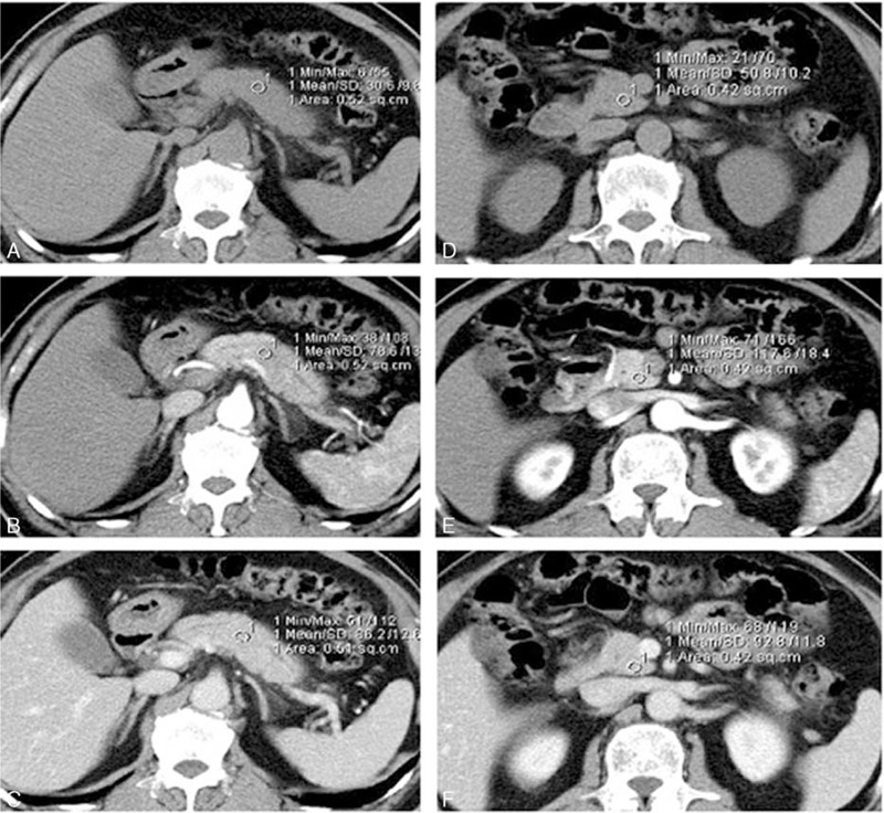
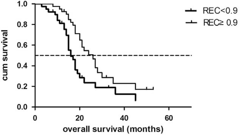
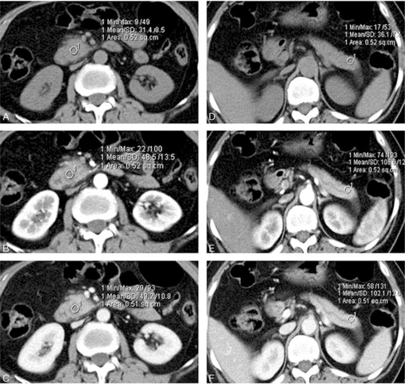
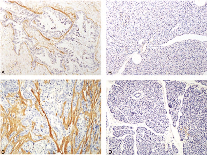
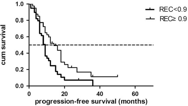
This study aimed to determine whether changes in contrast-enhanced computed tomography (CT) parameters could predict postsurgery overall and progression-free survival (PFS) in pancreatic cancer patients. Seventy-nine patients with a final pathological diagnosis of pancreatic adenocarcinoma were included in this study from June 2008 to August 2012. Dynamic contrast-enhanced (DCE) CT of tumors was obtained before curative-intent surgery. Absolute enhancement change (AEC) and relative enhancement change (REC) were evaluated on DCE-CT. PFS and overall survival (OS) were compared based on CT enhancement patterns. The markers of fibrogenic alpha-smooth muscle antigen (α-SMA) and periostin in tumor specimens were evaluated by immunohistochemical staining. The χ test was performed to determine whether CT enhancement patterns were associated with α-SMA-periostin expression levels (recorded as positive or negative). Lower REC (<0.9) was associated with shorter PFS (HR 0.51, 95% CI: 0.31-0.89) and OS (HR 0.44, 95% CI: 0.25-0.78). The α-SMA and periostin expression level were negatively correlated with REC (both P = 0). Among several CT enhancement parameters, REC was the best predictor of patient postsurgery survival. Low REC was associated with a short progression-free time and poor survival. The pathological studies suggested that REC might be a reflection of cancer fibrogenic potential.

摘要

本研究旨在确定对比增强计算机断层扫描(CT)参数的变化是否能够预测胰腺癌患者术后的总生存期和无进展生存期(PFS)。2008年6月至2012年8月,本研究纳入了79例最终病理诊断为胰腺腺癌的患者。在进行根治性手术前,对肿瘤进行动态对比增强(DCE)CT检查。在DCE-CT上评估绝对增强变化(AEC)和相对增强变化(REC)。根据CT增强模式比较PFS和总生存期(OS)。通过免疫组织化学染色评估肿瘤标本中促纤维化α平滑肌抗原(α-SMA)和骨膜蛋白的标志物。进行χ检验以确定CT增强模式是否与α-SMA-骨膜蛋白表达水平(记录为阳性或阴性)相关。较低的REC(<0.9)与较短的PFS(风险比[HR]0.51,95%置信区间[CI]:0.31-0.89)和OS(HR 0.44,95%CI:0.25-0.78)相关。α-SMA和骨膜蛋白表达水平与REC呈负相关(均P = 0)。在几个CT增强参数中,REC是患者术后生存的最佳预测指标。低REC与较短的无进展时间和较差的生存率相关。病理研究表明,REC可能反映了癌症的促纤维化潜能。

https://cdn.ncbi.nlm.nih.gov/pmc/blobs/4021/4748912/c5ebc9a5893a/medi-95-e2664-g012.jpg
https://cdn.ncbi.nlm.nih.gov/pmc/blobs/4021/4748912/f210127b0146/medi-95-e2664-g001.jpg
https://cdn.ncbi.nlm.nih.gov/pmc/blobs/4021/4748912/b3590e2978e3/medi-95-e2664-g002.jpg
https://cdn.ncbi.nlm.nih.gov/pmc/blobs/4021/4748912/0d3aae311a73/medi-95-e2664-g004.jpg
https://cdn.ncbi.nlm.nih.gov/pmc/blobs/4021/4748912/8b4ad5c2381a/medi-95-e2664-g010.jpg
https://cdn.ncbi.nlm.nih.gov/pmc/blobs/4021/4748912/c5ebc9a5893a/medi-95-e2664-g012.jpg

相似文献

1
CT Imaging Biomarkers Predict Clinical Outcomes After Pancreatic Cancer Surgery.CT成像生物标志物可预测胰腺癌手术后的临床结局。
Medicine (Baltimore). 2016 Feb;95(5):e2664. doi: 10.1097/MD.0000000000002664.
2
Small (≤ 20 mm) pancreatic adenocarcinomas: analysis of enhancement patterns and secondary signs with multiphasic multidetector CT.小(≤ 20mm)胰腺腺癌:多期多层 CT 增强模式和次要征象分析。
Radiology. 2011 May;259(2):442-52. doi: 10.1148/radiol.11101133. Epub 2011 Mar 15.
3
Visually isoattenuating pancreatic adenocarcinoma at dynamic-enhanced CT: frequency, clinical and pathologic characteristics, and diagnosis at imaging examinations.动态增强 CT 上呈等密度的胰腺腺癌:频率、临床和病理特征及影像学检查诊断。
Radiology. 2010 Oct;257(1):87-96. doi: 10.1148/radiol.10100015. Epub 2010 Aug 9.
4
Prognostic Value of FDG PET/CT-Derived Parameters in Pancreatic Adenocarcinoma at Initial PET/CT Staging.FDG PET/CT 衍生参数在初始 FDG PET/CT 分期时对胰腺腺癌的预后价值。
AJR Am J Roentgenol. 2015 May;204(5):1093-9. doi: 10.2214/AJR.14.13156.
5
Pancreatic cancer: value of dual-phase helical CT in assessing resectability.胰腺癌:双期螺旋CT在评估可切除性方面的价值
Radiology. 1998 Feb;206(2):373-8. doi: 10.1148/radiology.206.2.9457188.
6
Fibrous stroma and vascularity of pancreatic carcinoma: correlation with enhancement patterns on CT.胰腺癌的纤维性间质与血管分布:与CT增强模式的相关性
Abdom Imaging. 2010 Apr;35(2):172-80. doi: 10.1007/s00261-008-9460-0. Epub 2008 Sep 25.
7
Estimation of Extracellular Volume Fraction With Routine Multiphasic Pancreatic Computed Tomography to Predict the Survival of Patients With Stage IV Pancreatic Ductal Adenocarcinoma.利用常规多期胰腺计算机断层扫描估计细胞外体积分数以预测IV期胰腺导管腺癌患者的生存情况
Pancreas. 2019 Nov/Dec;48(10):1360-1366. doi: 10.1097/MPA.0000000000001427.
8
Dynamic contrast-enhanced MR imaging and dual-phase helical CT in the preoperative assessment of suspected pancreatic cancer: a comparative study with receiver operating characteristic analysis.动态对比增强磁共振成像和双期螺旋CT在疑似胰腺癌术前评估中的应用:一项采用受试者操作特征分析的对比研究
AJR Am J Roentgenol. 1999 Sep;173(3):583-90. doi: 10.2214/ajr.173.3.10470884.
9
Enhancement patterns of pancreatic adenocarcinoma on conventional dynamic multi-detector row CT: correlation with angiogenesis and fibrosis.胰腺腺癌在传统动态多排螺旋CT上的强化模式:与血管生成和纤维化的相关性
World J Gastroenterol. 2009 Jul 7;15(25):3114-21. doi: 10.3748/wjg.15.3114.
10
Preoperative CT in patients with surgically resectable pancreatic adenocarcinoma: does the time interval between CT and surgery affect survival?术前 CT 检查在可手术切除的胰腺腺癌患者中的应用:CT 检查与手术之间的时间间隔是否会影响生存?
Abdom Radiol (NY). 2018 Mar;43(3):620-628. doi: 10.1007/s00261-017-1254-9.

引用本文的文献

1
Preoperative Prognostic Factors in Resectable Pancreatic Cancer: State of the Art and Prospects.可切除胰腺癌的术前预后因素:现状与展望
Ann Surg Oncol. 2025 Jun;32(6):4117-4127. doi: 10.1245/s10434-025-17062-w. Epub 2025 Mar 17.
2
Performance of different CT enhancement quantification methods as predictors of pancreatic cancer recurrence after upfront surgery.不同 CT 增强量化方法在预测术前手术治疗后胰腺癌复发中的表现。
Sci Rep. 2024 Aug 26;14(1):19783. doi: 10.1038/s41598-024-70441-3.
3
Rim Enhancement on Contrast-Enhanced CT as a Predictor of Prognosis in Patients with Pancreatic Ductal Adenocarcinoma.

本文引用的文献

1
Pancreatic neuroendocrine tumors: Correlation between the contrast-enhanced computed tomography features and the pathological tumor grade.胰腺神经内分泌肿瘤:增强计算机断层扫描特征与肿瘤病理分级之间的相关性
Eur J Radiol. 2015 Aug;84(8):1436-1443. doi: 10.1016/j.ejrad.2015.05.005. Epub 2015 May 14.
2
Detection of small (≤ 2 cm) pancreatic adenocarcinoma and surrounding parenchyma: correlations between enhancement patterns at triphasic MDCT and histologic features.小(≤2 cm)胰腺腺癌及其周围实质的检测:多期多层螺旋CT增强模式与组织学特征的相关性
BMC Gastroenterol. 2014 Jan 21;14:16. doi: 10.1186/1471-230X-14-16.
3
对比增强CT上的边缘强化作为胰腺导管腺癌患者预后的预测指标
Diagnostics (Basel). 2024 Apr 9;14(8):782. doi: 10.3390/diagnostics14080782.
4
Radiomics analysis with three-dimensional and two-dimensional segmentation to predict survival outcomes in pancreatic cancer.采用三维和二维分割的放射组学分析预测胰腺癌的生存结果。
World J Radiol. 2023 Nov 28;15(11):304-314. doi: 10.4329/wjr.v15.i11.304.
5
Predicting the Efficacy of Neoadjuvant Chemotherapy for Pancreatic Cancer Using Deep Learning of Contrast-Enhanced Ultrasound Videos.使用对比增强超声视频的深度学习预测胰腺癌新辅助化疗的疗效
Diagnostics (Basel). 2023 Jun 27;13(13):2183. doi: 10.3390/diagnostics13132183.
6
Prognostic value of preoperative enhanced computed tomography as a quantitative imaging biomarker in pancreatic cancer.术前增强 CT 作为一种定量成像生物标志物对胰腺癌的预后价值。
World J Gastroenterol. 2022 Jun 14;28(22):2468-2481. doi: 10.3748/wjg.v28.i22.2468.
7
A predictive model for recurrence after upfront surgery in patients with resectable pancreatic ductal adenocarcinoma (PDAC) by using preoperative clinical data and CT characteristics.术前临床数据和 CT 特征预测可切除胰腺导管腺癌(PDAC)患者 upfront 手术后复发的模型。
BMC Med Imaging. 2022 Jul 3;22(1):116. doi: 10.1186/s12880-022-00823-4.
8
Basic Pancreatic Lesions: Radiologic-pathologic Correlation.胰腺基本病变:放射学与病理学的相关性
J Transl Int Med. 2022 Apr 2;10(1):18-27. doi: 10.2478/jtim-2022-0003. eCollection 2022 Mar.
9
Correlation Between Enhancement Patterns on Transabdominal Ultrasound and Survival for Pancreatic Ductal Adenocarcinoma.经腹超声增强模式与胰腺导管腺癌生存率的相关性
Cancer Manag Res. 2021 Aug 31;13:6823-6832. doi: 10.2147/CMAR.S307079. eCollection 2021.
10
Predictive Modeling for Voxel-Based Quantification of Imaging-Based Subtypes of Pancreatic Ductal Adenocarcinoma (PDAC): A Multi-Institutional Study.基于体素的胰腺导管腺癌(PDAC)影像亚型定量的预测建模:一项多机构研究
Cancers (Basel). 2020 Dec 5;12(12):3656. doi: 10.3390/cancers12123656.
Contrast-enhanced CT and diffusion-weighted MR imaging: performance as a prognostic factor in patients with pancreatic ductal adenocarcinoma.
对比增强CT和扩散加权磁共振成像:作为胰腺导管腺癌患者预后因素的表现
Eur J Radiol. 2014 Apr;83(4):612-9. doi: 10.1016/j.ejrad.2013.12.016. Epub 2013 Dec 27.
4
Prognostic estimator of survival for patients with localized and extended pancreatic ductal adenocarcinoma.局限性和广泛性胰腺导管腺癌患者生存的预后评估指标
Cancer Inform. 2013 Mar 18;12:103-14. doi: 10.4137/CIN.S11496. Print 2013.
5
The role of pancreatic stellate cells in pancreatic cancer.胰腺星状细胞在胰腺癌中的作用。
Pancreatology. 2013 Mar-Apr;13(2):106-9. doi: 10.1016/j.pan.2013.01.008. Epub 2013 Feb 5.
6
Pancreatic cancer-associated stellate cells promote differentiation of myeloid-derived suppressor cells in a STAT3-dependent manner.胰腺癌相关星型细胞STAT3 依赖性促进髓源抑制细胞分化。
Cancer Res. 2013 May 15;73(10):3007-18. doi: 10.1158/0008-5472.CAN-12-4601. Epub 2013 Mar 20.
7
Clinical features and treatment outcome of borderline resectable pancreatic head/body cancer: a multi-institutional survey by the Japanese Society of Pancreatic Surgery.可切除边缘性胰头/体部癌的临床特征及治疗结果:日本胰腺外科学会的多机构调查
J Hepatobiliary Pancreat Sci. 2013 Aug;20(6):601-10. doi: 10.1007/s00534-013-0595-1.
8
Borderline resectable pancreatic cancer: need for standardization and methods for optimal clinical trial design.交界可切除胰腺癌:标准化的必要性和优化临床试验设计的方法。
Ann Surg Oncol. 2013 Aug;20(8):2787-95. doi: 10.1245/s10434-013-2886-9. Epub 2013 Feb 23.
9
Pirfenidone inhibits pancreatic cancer desmoplasia by regulating stellate cells.吡非尼酮通过调控星型细胞抑制胰腺癌基质纤维化。
Cancer Res. 2013 Apr 1;73(7):2345-56. doi: 10.1158/0008-5472.CAN-12-3180. Epub 2013 Jan 24.
10
Pancreatic neuroendocrine neoplasm: correlation between computed tomography enhancement patterns and prognostic factors of surgical and endoscopic ultrasound-guided fine-needle aspiration biopsy specimens.胰腺神经内分泌肿瘤:计算机断层扫描增强模式与手术及内镜超声引导下细针穿刺活检标本预后因素的相关性
Abdom Imaging. 2013 Apr;38(2):358-66. doi: 10.1007/s00261-012-9953-8.