• 文献检索
  • 文档翻译
  • 深度研究
  • 学术资讯
  • Suppr Zotero 插件Zotero 插件
  • 邀请有礼
  • 套餐&价格
  • 历史记录
应用&插件
Suppr Zotero 插件Zotero 插件浏览器插件Mac 客户端Windows 客户端微信小程序
定价
高级版会员购买积分包购买API积分包
服务
文献检索文档翻译深度研究API 文档MCP 服务
关于我们
关于 Suppr公司介绍联系我们用户协议隐私条款
关注我们

Suppr 超能文献

核心技术专利:CN118964589B侵权必究
粤ICP备2023148730 号-1Suppr @ 2026

文献检索

告别复杂PubMed语法,用中文像聊天一样搜索,搜遍4000万医学文献。AI智能推荐,让科研检索更轻松。

立即免费搜索

文件翻译

保留排版,准确专业,支持PDF/Word/PPT等文件格式,支持 12+语言互译。

免费翻译文档

深度研究

AI帮你快速写综述,25分钟生成高质量综述,智能提取关键信息,辅助科研写作。

立即免费体验

一种简单、快速且可重复的用于评估左心室舒张功能分级的超声心动图方法。

A simple, fast and reproducible echocardiographic approach to grade left ventricular diastolic function.

作者信息

van Dalen Bas M, Strachinaru Mihai, van der Swaluw Julio, Geleijnse Marcel L

机构信息

Department of Cardiology, The Thoraxcenter, Erasmus University Medical Center, 's-Gravendijkwal 230, Room Bd412, 3015 CE, Rotterdam, The Netherlands.

Department of Cardiology, Sint Franciscus Gasthuis, Rotterdam, The Netherlands.

出版信息

Int J Cardiovasc Imaging. 2016 May;32(5):743-52. doi: 10.1007/s10554-015-0832-6. Epub 2016 Feb 3.

DOI:10.1007/s10554-015-0832-6
PMID:26847461
原文链接:https://pmc.ncbi.nlm.nih.gov/articles/PMC4853445/
Abstract

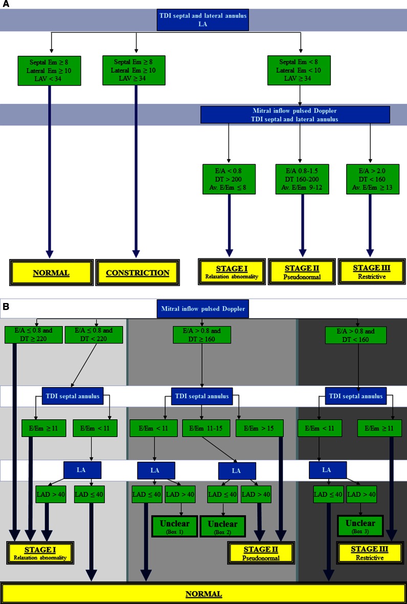
The American Society of Echocardiography and European Association of Echocardiography (ASE/EAE) have published an algorithm for the grading of diastolic function. However, the ability to use this algorithm effectively in daily clinical practice has not been investigated. We hypothesized that in some patients it may be difficult to grade diastolic dysfunction with this scheme, since there may be discrepancies in the assessed parameters. The aim of the current study was to test the feasibility of the ASE/EAE algorithm and to compare this with a new Thoraxcenter (TXC) algorithm. The ASE/EAE and TXC algorithms were applied to 200 patients. The ASE/EAE algorithm starts with assessment of diastolic myocardial wall velocities and left atrial (LA) volumes with subsequent assessment of E/A ratio, E-wave deceleration time and pulmonary venous flow. The TXC algorithm reverses these steps, uses LA dimension instead of volume and does not include a Valsalva manoeuvre and pulmonary venous flow. Due to inconsistencies between diastolic myocardial wall velocities and LA volumes and a not covered E/A ratio in the range of 1.5-2 it was not possible to classify 48 % of patients with the ASE/EAE algorithm, as opposed to only 10 % by the TXC algorithm. LA volume was always needed in the ASE/EAE algorithm. In only 64 % of patients LA size was necessary by the TXC algorithm. When LA volume would have been used instead of LA dimension, grading of LV diastolic function would have been different in only 2 % of patients without apparent improvement. Assessment of LA dimension was considerably faster than LA volume. The TXC algorithm to grade LV diastolic dysfunction was compared to the ASE/EAE algorithm simpler, faster, better reproducible and yields a higher diagnostic outcome.

摘要

美国超声心动图学会和欧洲超声心动图协会(ASE/EAE)已发布了一种舒张功能分级算法。然而,在日常临床实践中有效使用该算法的能力尚未得到研究。我们推测,在某些患者中,使用该方案对舒张功能障碍进行分级可能存在困难,因为评估参数可能存在差异。本研究的目的是测试ASE/EAE算法的可行性,并将其与一种新的Thoraxcenter(TXC)算法进行比较。将ASE/EAE和TXC算法应用于200例患者。ASE/EAE算法首先评估舒张期心肌壁速度和左心房(LA)容积,随后评估E/A比值、E波减速时间和肺静脉血流。TXC算法颠倒了这些步骤,使用LA内径而非容积,且不包括瓦尔萨尔瓦动作和肺静脉血流。由于舒张期心肌壁速度与LA容积之间存在不一致,且E/A比值在1.5 - 2范围内未涵盖,使用ASE/EAE算法无法对48%的患者进行分类,而TXC算法仅为10%。ASE/EAE算法始终需要LA容积。TXC算法仅在64%的患者中需要LA大小。如果使用LA容积而非LA内径,在仅2%的患者中左心室舒张功能分级会有所不同,且无明显改善。LA内径的评估比LA容积快得多。与ASE/EAE算法相比,TXC算法对左心室舒张功能障碍进行分级更简单、更快、重复性更好且诊断结果更高。

https://cdn.ncbi.nlm.nih.gov/pmc/blobs/bb70/4853445/6d351f5fe17d/10554_2015_832_Fig1_HTML.jpg

相似文献

1
A simple, fast and reproducible echocardiographic approach to grade left ventricular diastolic function.一种简单、快速且可重复的用于评估左心室舒张功能分级的超声心动图方法。
Int J Cardiovasc Imaging. 2016 May;32(5):743-52. doi: 10.1007/s10554-015-0832-6. Epub 2016 Feb 3.
2
Transthoracic 3D Echocardiographic Left Heart Chamber Quantification Using an Automated Adaptive Analytics Algorithm.经胸三维超声心动图左心腔定量分析的自动自适应分析算法。
JACC Cardiovasc Imaging. 2016 Jul;9(7):769-782. doi: 10.1016/j.jcmg.2015.12.020. Epub 2016 Jun 15.
3
A real-time three-dimensional echocardiographic quantitative analysis of left atrial function in left ventricular diastolic dysfunction.左心室舒张功能障碍时左心房功能的实时三维超声心动图定量分析
Am J Cardiol. 2008 Oct 15;102(8):1097-102. doi: 10.1016/j.amjcard.2008.05.067. Epub 2008 Aug 5.
4
How to incorporate left atrial strain in the diagnostic algorithm of left ventricular diastolic dysfunction.如何将左心房应变纳入左心室舒张功能障碍的诊断算法中。
Int J Cardiovasc Imaging. 2021 Mar;37(3):945-951. doi: 10.1007/s10554-020-02070-6. Epub 2020 Oct 31.
5
[The Evaluation of Left Ventricular Diastolic Function by Echocardiography in Patients With Preserved Ejection Fraction].[超声心动图评估射血分数保留患者的左心室舒张功能]
Kardiologiia. 2017 Feb;57(2):40-45.
6
Association of left atrial reservoir function with left atrial structural remodeling related to left ventricular dysfunction in asymptomatic patients with hypertension: evaluation by two-dimensional speckle-tracking echocardiography.二维斑点追踪超声心动图评价无症状高血压患者左心房储器功能与左心室功能障碍相关左心房结构重构的关系。
Clin Exp Hypertens. 2015;37(2):155-65. doi: 10.3109/10641963.2014.933962. Epub 2014 Jul 22.
7
Classification of left ventricular diastolic function using American Society of Echocardiography Guidelines: agreement among echocardiographers.使用美国超声心动图学会指南对左心室舒张功能进行分类:超声心动图医生之间的一致性。
Echocardiography. 2013 Oct;30(9):1022-31. doi: 10.1111/echo.12185. Epub 2013 Apr 1.
8
Strain rate imaging for noninvasive functional quantification of the left atrium: comparative studies in controls and patients with atrial fibrillation.应变率成像用于左心房无创功能定量分析:对照者与心房颤动患者的对比研究
J Am Soc Echocardiogr. 2005 Jul;18(7):729-36. doi: 10.1016/j.echo.2004.12.005.
9
Redefining diastolic dysfunction grading: combination of E/A ≤0.75 and deceleration time >140 ms and E/ε' ≥10.重新定义舒张功能障碍分级:E/A≤0.75 与减速时间>140ms 联合,以及 E/ε'≥10。
JACC Cardiovasc Imaging. 2014 Aug;7(8):749-58. doi: 10.1016/j.jcmg.2014.05.002. Epub 2014 Jul 16.
10
Impact of Left Ventricular Diastolic Property on Left Atrial Function from Simultaneous Left Atrial and Ventricular Three-Dimensional Echocardiographic Volume Measurement.基于同步左心房和心室三维超声心动图容积测量评估左心室舒张特性对左心房功能的影响
Am J Cardiol. 2017 May 15;119(10):1687-1693. doi: 10.1016/j.amjcard.2017.02.002. Epub 2017 Feb 28.

引用本文的文献

1
Automated algorithms in diastology: how to move forward?舒张学中的自动化算法:如何向前发展?
Int J Cardiovasc Imaging. 2022 May;38(5):975-977. doi: 10.1007/s10554-021-02505-8. Epub 2022 Feb 8.
2
Can echocardiographic assessment of diastolic function be automated?舒张功能的超声心动图评估能否实现自动化?
Int J Cardiovasc Imaging. 2022 May;38(5):965-974. doi: 10.1007/s10554-021-02488-6. Epub 2021 Dec 9.
3
Relationship of left ventricular and atrial dimensions with moderate to severe left ventricular diastolic dysfunction (grade II and above).

本文引用的文献

1
Comparison of the reliability of E/E' to estimate pulmonary capillary wedge pressure in heart failure patients with preserved ejection fraction versus those with reduced ejection fraction.射血分数保留的心力衰竭患者与射血分数降低的心力衰竭患者中,E/E' 用于估计肺毛细血管楔压的可靠性比较。
Int J Cardiovasc Imaging. 2015 Dec;31(8):1497-502. doi: 10.1007/s10554-015-0718-7. Epub 2015 Jul 28.
2
Relation between E/e' ratio and NT-proBNP levels in elderly patients with symptomatic severe aortic stenosis.有症状的重度主动脉瓣狭窄老年患者中E/e'比值与N末端B型利钠肽原水平的关系
Cardiovasc Ultrasound. 2015 Jun 26;13:29. doi: 10.1186/s12947-015-0021-8.
3
左心室和心房容积与中度至重度左心室舒张功能障碍(II 级及以上)的关系。
Afr Health Sci. 2020 Dec;20(4):1749-1753. doi: 10.4314/ahs.v20i4.27.
4
A simplified definition of diastolic function in sepsis, compared against standard definitions.与标准定义相比,脓毒症中舒张功能的简化定义。
J Intensive Care. 2019 Feb 20;7:14. doi: 10.1186/s40560-019-0367-3. eCollection 2019.
5
Early detection of left ventricular diastolic dysfunction using conventional and speckle tracking echocardiography in a large animal model of metabolic dysfunction.在代谢功能障碍的大型动物模型中,使用传统超声心动图和斑点追踪超声心动图早期检测左心室舒张功能障碍。
Int J Cardiovasc Imaging. 2018 May;34(5):743-749. doi: 10.1007/s10554-017-1287-8. Epub 2017 Dec 12.
6
Cardiovascular imaging 2016 in the International Journal of Cardiovascular Imaging.《国际心血管影像杂志》2016年心血管成像相关内容
Int J Cardiovasc Imaging. 2017 Jun;33(6):761-770. doi: 10.1007/s10554-017-1111-5.
Recommendations for cardiac chamber quantification by echocardiography in adults: an update from the American Society of Echocardiography and the European Association of Cardiovascular Imaging.
超声心动图成人左心室容量和射血分数测量:美国超声心动图学会和欧洲心血管影像协会的更新建议。
J Am Soc Echocardiogr. 2015 Jan;28(1):1-39.e14. doi: 10.1016/j.echo.2014.10.003.
4
Redefining diastolic dysfunction grading: combination of E/A ≤0.75 and deceleration time >140 ms and E/ε' ≥10.重新定义舒张功能障碍分级:E/A≤0.75 与减速时间>140ms 联合,以及 E/ε'≥10。
JACC Cardiovasc Imaging. 2014 Aug;7(8):749-58. doi: 10.1016/j.jcmg.2014.05.002. Epub 2014 Jul 16.
5
Incremental prognostic value of diastolic dysfunction in low risk patients undergoing echocardiography: beyond Framingham score.超声心动图检查低危患者舒张功能障碍的预后价值递增:超越弗雷明汉评分。
Int J Cardiovasc Imaging. 2013 Oct;29(7):1441-50. doi: 10.1007/s10554-013-0246-2. Epub 2013 Jul 11.
6
The epidemiology of heart failure, based on data for 2.1 million inhabitants in Sweden.心力衰竭的流行病学,基于瑞典 210 万居民的数据。
Eur J Heart Fail. 2013 Sep;15(9):995-1002. doi: 10.1093/eurjhf/hft064. Epub 2013 May 3.
7
Echo-Doppler assessment of diastole: flow, function and haemodynamics.
Heart. 2013 Jan;99(1):55-64. doi: 10.1136/heartjnl-2012-301961.
8
Indexed left atrial volume is superior to left atrial diameter in predicting nonvalvular atrial fibrillation recurrence after successful cardioversion: a prospective study.在预测成功复律后非瓣膜性心房颤动复发方面,左心房容积指数优于左心房直径:一项前瞻性研究。
Echocardiography. 2012 Mar;29(3):276-84. doi: 10.1111/j.1540-8175.2011.01580.x. Epub 2011 Nov 18.
9
Usefulness of left atrial volume versus diameter to assess thromboembolic risk in mitral stenosis.左心房容积与直径评估二尖瓣狭窄血栓栓塞风险的作用。
Am J Cardiol. 2010 Oct 15;106(8):1152-6. doi: 10.1016/j.amjcard.2010.06.024.
10
Recommendations for the evaluation of left ventricular diastolic function by echocardiography.超声心动图评估左心室舒张功能的建议。
J Am Soc Echocardiogr. 2009 Feb;22(2):107-33. doi: 10.1016/j.echo.2008.11.023.