Kijanka Marta M, van Brussel Aram S A, van der Wall Elsken, Mali Willem P T M, van Diest Paul J, van Bergen En Henegouwen Paul M P, Oliveira Sabrina
Division of Cell Biology, Department of Biology, Science Faculty, Utrecht University, Utrecht, The Netherlands.
Department of Pathology, University Medical Center Utrecht, Utrecht, The Netherlands.
EJNMMI Res. 2016 Dec;6(1):14. doi: 10.1186/s13550-016-0166-y. Epub 2016 Feb 10.
Optical molecular imaging is an emerging novel technology with applications in the diagnosis of cancer and assistance in image-guided surgery. A high tumour-to-background (T/B) ratio is crucial for successful imaging, which strongly depends on tumour-specific probes that rapidly accumulate in the tumour, while non-bound probes are rapidly cleared. Here, using pre-invasive breast cancer as a model, we investigate whether the use of combinations of probes with different target specificities results in higher T/B ratios and whether dual-spectral imaging leads to improvements in tumour characterization.
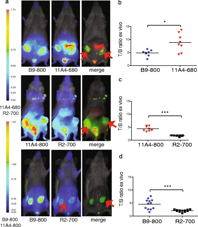
We performed optical molecular imaging of an orthotopic breast cancer model mimicking ductal carcinoma in situ (DCIS). A combination of carbonic anhydrase IX (CAIX)- and human epidermal growth factor receptor 2 (HER2)-specific variable domains of the heavy chain from heavy-chain antibodies (VHHs) was conjugated either to the same fluorophore (IRDye800CW) to evaluate T/B ratios or to different fluorophores (IRDye800CW, IRDye680RD or IRDye700DX) to analyse the expression of CAIX and HER2 simultaneously through dual-fluorescence detection. These experiments were performed non-invasively in vivo, in a mimicked intra-operative setting, and ex vivo on tumour sections.
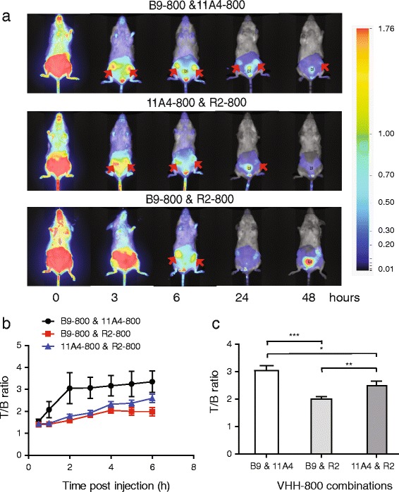
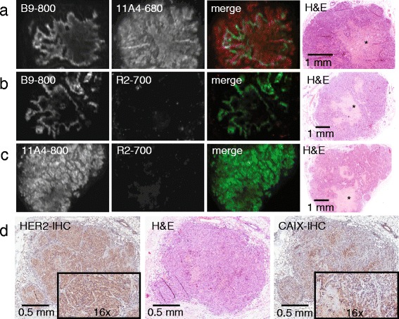
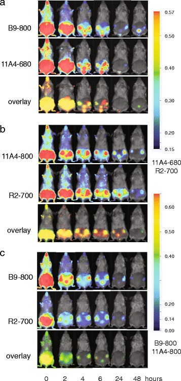
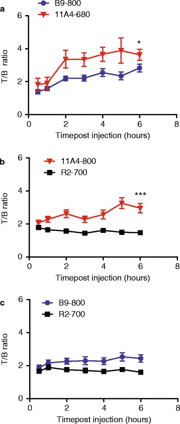
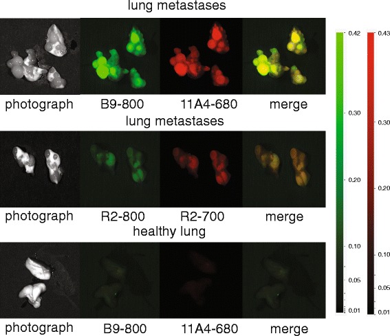
Application of the CAIX- and HER2-specific VHH combination resulted in an increase of the T/B ratio, as compared to T/B ratios obtained from each of these single VHHs together with an irrelevant VHH. This dual tumour marker-specific VHH combination also enabled the detection of small metastases in the lung. Furthermore, dual-spectral imaging enabled the assessment of the expression status of both CAIX and HER2 in a mimicked intra-operative setting, as well as on tumour sections, which was confirmed by immunohistochemistry.
These results establish the feasibility of the use of VHH 'cocktails' to increase T/B ratios and improve early detection of heterogeneous tumours and the use of multispectral molecular imaging to facilitate the assessment of the target expression status of tumours and metastases, both invasive or non-invasively.
光学分子成像技术是一项新兴的新技术,可应用于癌症诊断及辅助图像引导手术。高肿瘤与背景(T/B)比对于成功成像至关重要,这很大程度上依赖于能在肿瘤中快速积聚的肿瘤特异性探针,同时未结合的探针能被迅速清除。在此,我们以侵袭前乳腺癌为模型,研究使用具有不同靶点特异性的探针组合是否会导致更高的T/B比,以及双光谱成像是否能改善肿瘤特征描述。
我们对模拟原位导管癌(DCIS)的原位乳腺癌模型进行了光学分子成像。将碳酸酐酶IX(CAIX)特异性和人表皮生长因子受体2(HER2)特异性的重链抗体可变区(VHHs)与同一荧光团(IRDye800CW)偶联以评估T/B比,或与不同荧光团(IRDye800CW、IRDye680RD或IRDye700DX)偶联,通过双荧光检测同时分析CAIX和HER2的表达。这些实验在体内以模拟术中环境进行非侵入性操作,并在肿瘤切片上进行离体操作。
与单独使用这些单一VHH以及无关VHH所获得的T/B比相比,应用CAIX和HER2特异性VHH组合可使T/B比增加。这种双肿瘤标志物特异性VHH组合还能够检测出肺部的小转移灶。此外,双光谱成像能够在模拟术中环境以及肿瘤切片上评估CAIX和HER2的表达状态,免疫组织化学证实了这一点。
这些结果证实了使用VHH“鸡尾酒”提高T/B比并改善异质性肿瘤早期检测的可行性,以及使用多光谱分子成像促进对肿瘤和转移灶的靶点表达状态进行评估的可行性,包括侵入性和非侵入性评估。