Andjelkov Nenad, Hamberg Hans, Bjellerup Per
Department of Orthopaedics, Västmanland County Hospital, Västerås, Sweden.
Centre for Clinical Research, Uppsala University, Västmanland County Hospital, Västerås, Sweden.
J Orthop Surg Res. 2016 Feb 16;11:23. doi: 10.1186/s13018-016-0355-4.
Commercially available fibrin is routinely being used as both a matrix in certain cartilage repair techniques and a method for scaffold fixation. Chondrocytes from non-digested particulated cartilage fragments are proposed as a possible source for new cartilage tissue formation in some operative techniques. The goal of this study was to test that chondrocytes from particulated articular cartilage embedded in fibrin have an active role in the process of cartilage repair, as well as if commercially available fibrin should be used as a suitable matrix.
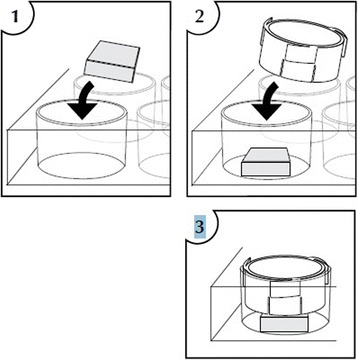
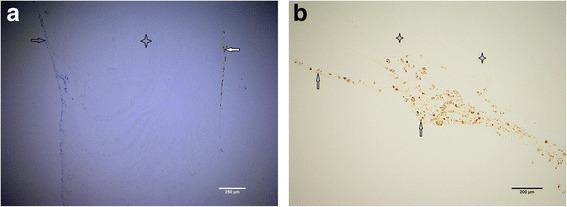
Articular cartilage was obtained from patients undergoing total knee replacement surgery. The biopsies were particulated in small, 1-2-mm(3) pieces and embedded in fibrin. Two groups were compared in our study, particulated articular cartilage with and without collagenase treatment. The specimens were analyzed by optical microscopy after 2-5 weeks of cultivation in a special construct embedded in a cell culture medium containing particulated cartilage embedded in fibrin in the upper phase and cancellous bone in the lower phase under the perforated nylon membrane.
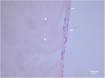
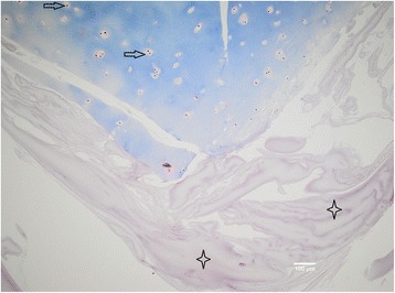
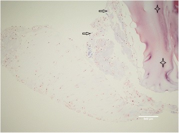
None of the biopsies taken from four different patients showed the outgrowth of chondrocytes or bone marrow-originated cells into the fibrin matrix in our experimental model.
It has been shown in our experimental model in vitro little to support the theory that articular chondrocytes from particulated articular cartilage embedded in fibrin have an active role in cartilage repair in its early stage.
市售纤维蛋白通常既用作某些软骨修复技术中的基质,也用作支架固定方法。在一些手术技术中,来自未消化的颗粒状软骨碎片的软骨细胞被认为是新软骨组织形成的可能来源。本研究的目的是测试包埋在纤维蛋白中的颗粒状关节软骨中的软骨细胞在软骨修复过程中是否发挥积极作用,以及市售纤维蛋白是否应作为合适的基质使用。
从接受全膝关节置换手术的患者获取关节软骨。将活检组织切成1 - 2立方毫米的小块并包埋在纤维蛋白中。在本研究中比较了两组,即经过和未经过胶原酶处理的颗粒状关节软骨。将标本置于一种特殊构建体中,该构建体嵌入含有上层为包埋在纤维蛋白中的颗粒状软骨、下层为松质骨且中间有穿孔尼龙膜的细胞培养基中培养2 - 5周后,通过光学显微镜进行分析。
在我们的实验模型中,取自四名不同患者的活检组织均未显示软骨细胞或骨髓来源细胞向纤维蛋白基质中生长。
在我们的体外实验模型中,几乎没有证据支持包埋在纤维蛋白中的颗粒状关节软骨中的关节软骨细胞在早期软骨修复中发挥积极作用这一理论。