Arora Abishek, Bhagat Neeta
Amity Institute of Biotechnology, Amity University Uttar Pradesh, Noida 201303, India.
Amity Institute of Biotechnology, Amity University Uttar Pradesh, Room No. 312, J3 Block, III Floor, Noida 201303, India.
Int J Biomed Imaging. 2016;2016:7462014. doi: 10.1155/2016/7462014. Epub 2016 Jan 10.
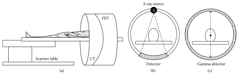
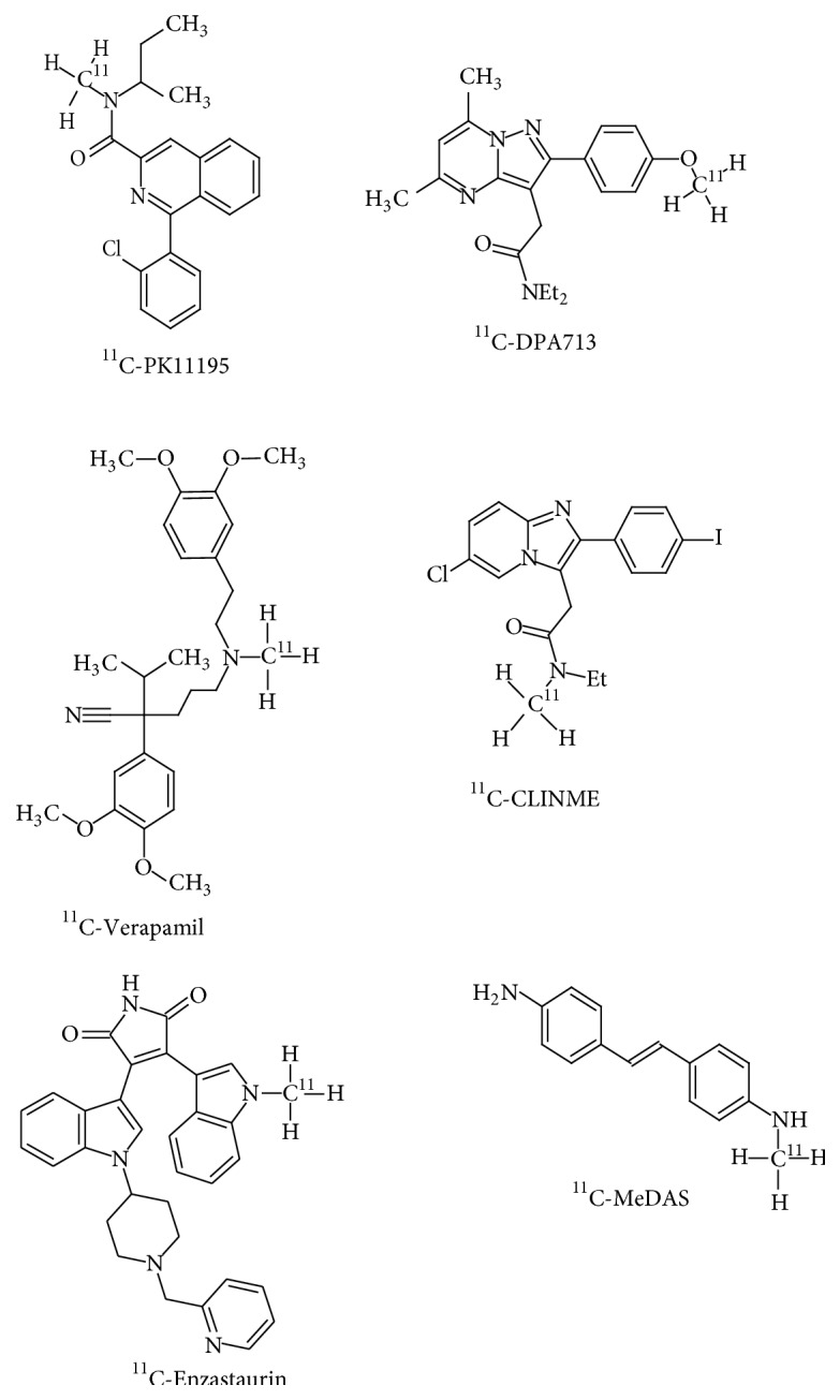
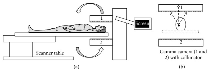
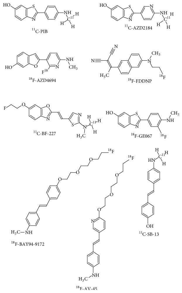
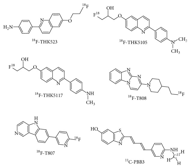
Alzheimer's disease is a complex neurodegenerative disease affecting millions of individuals worldwide. Earlier it was diagnosed only via clinical assessments and confirmed by postmortem brain histopathology. The development of validated biomarkers for Alzheimer's disease has given impetus to improve diagnostics and accelerate the development of new therapies. Functional imaging like positron emission tomography (PET), single photon emission computed tomography (SPECT), functional magnetic resonance imaging (fMRI), and proton magnetic resonance spectroscopy provides a means of detecting and characterising the regional changes in brain blood flow, metabolism, and receptor binding sites that are associated with Alzheimer's disease. Multimodal neuroimaging techniques have indicated changes in brain structure and metabolic activity, and an array of neurochemical variations that are associated with neurodegenerative diseases. Radiotracer-based PET and SPECT potentially provide sensitive, accurate methods for the early detection of disease. This paper presents a review of neuroimaging modalities like PET, SPECT, and selected imaging biomarkers/tracers used for the early diagnosis of AD. Neuroimaging with such biomarkers and tracers could achieve a much higher diagnostic accuracy for AD and related disorders in the future.
阿尔茨海默病是一种复杂的神经退行性疾病,影响着全球数百万人。早期,它只能通过临床评估进行诊断,并通过死后脑组织病理学加以证实。阿尔茨海默病有效生物标志物的开发推动了诊断的改进,并加速了新疗法的研发。正电子发射断层扫描(PET)、单光子发射计算机断层扫描(SPECT)、功能磁共振成像(fMRI)和质子磁共振波谱等功能成像提供了一种检测和表征与阿尔茨海默病相关的脑血流、代谢和受体结合位点区域变化的方法。多模态神经成像技术已显示出与神经退行性疾病相关的脑结构和代谢活动变化,以及一系列神经化学变化。基于放射性示踪剂的PET和SPECT有可能为疾病的早期检测提供灵敏、准确的方法。本文综述了PET、SPECT等神经成像模态以及用于阿尔茨海默病早期诊断的选定成像生物标志物/示踪剂。未来,使用此类生物标志物和示踪剂的神经成像可能会大大提高阿尔茨海默病及相关疾病的诊断准确性。