Zhao Fang, Li Xichun, Jin Liang, Zhang Fan, Inoue Masayuki, Yu Boyang, Cao Zhengyu
State Key Laboratory of Natural Medicines, China Pharmaceutical University, Nanjing 211198, China.
Jiangsu Provincial Key laboratory for TCM Evaluation and Translational Development, China Pharmaceutical University, Nanjing 211198, China.
Mar Drugs. 2016 Feb 16;14(2):36. doi: 10.3390/md14020036.
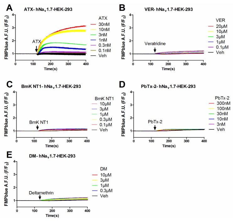
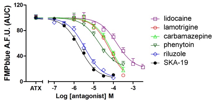
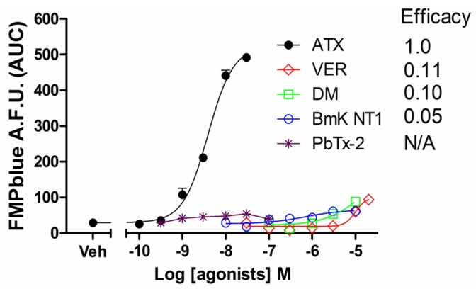
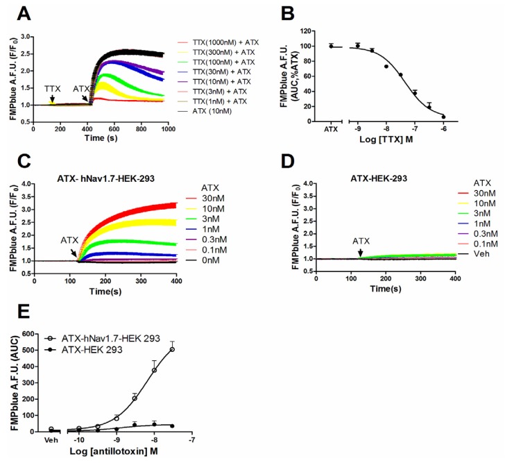
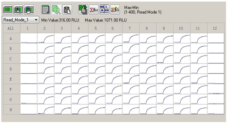
Voltage-gated sodium channels (VGSCs) are responsible for the generation of the action potential. Among nine classified VGSC subtypes (Nav1.1-Nav1.9), Nav1.7 is primarily expressed in the sensory neurons, contributing to the nociception transmission. Therefore Nav1.7 becomes a promising target for analgesic drug development. In this study, we compared the influence of an array of VGSC agonists including veratridine, BmK NT1, brevetoxin-2, deltamethrin and antillatoxin (ATX) on membrane depolarization which was detected by Fluorescence Imaging Plate Reader (FLIPR) membrane potential (FMP) blue dye. In HEK-293 cells heterologously expressing hNav1.7 α-subunit, ATX produced a robust membrane depolarization with an EC50 value of 7.8 ± 2.9 nM whereas veratridine, BmK NT1, and deltamethrin produced marginal response. Brevetoxin-2 was without effect on membrane potential change. The ATX response was completely inhibited by tetrodotoxin suggesting that the ATX response was solely derived from hNav1.7 activation, which was consistent with the results where ATX produced a negligible response in null HEK-293 cells. Six VGSC antagonists including lidocaine, lamotrigine, phenytoin, carbamazepine, riluzole, and 2-amino-6-trifluoromethylthiobenzothiazole all concentration-dependently inhibited ATX response with IC50 values comparable to that reported from patch-clamp experiments. Considered together, we demonstrate that ATX is a unique efficacious hNav1.7 activator which offers a useful probe to develop a rapid throughput screening assay to identify hNav1.7 antagonists.
电压门控钠通道(VGSCs)负责动作电位的产生。在九种分类的VGSC亚型(Nav1.1 - Nav1.9)中,Nav1.7主要在感觉神经元中表达,有助于伤害性感受的传递。因此,Nav1.7成为镇痛药开发的一个有前景的靶点。在本研究中,我们比较了一系列VGSC激动剂,包括藜芦碱、BmK NT1、短裸甲藻毒素 - 2、溴氰菊酯和海葵毒素(ATX)对膜去极化的影响,膜去极化通过荧光成像微孔板读数仪(FLIPR)膜电位(FMP)蓝色染料进行检测。在异源表达hNav1.7 α亚基的HEK - 293细胞中,ATX产生了强烈的膜去极化,EC50值为7.8±2.9 nM,而藜芦碱、BmK NT1和溴氰菊酯产生的反应微弱。短裸甲藻毒素 - 2对膜电位变化没有影响。河豚毒素完全抑制了ATX反应,表明ATX反应仅源于hNav1.7的激活,这与ATX在空HEK - 293细胞中产生可忽略不计的反应的结果一致。六种VGSC拮抗剂,包括利多卡因、拉莫三嗪、苯妥英、卡马西平、利鲁唑和2 - 氨基 - 6 - 三氟甲基硫代苯并噻唑,均浓度依赖性地抑制ATX反应,IC50值与膜片钳实验报道的值相当。综合考虑,我们证明ATX是一种独特有效的hNav1.7激活剂,它为开发一种快速通量筛选测定法以鉴定hNav1.7拮抗剂提供了一种有用的探针。