Park Ji Hye, Kim Chung Kwon, Lee Sang Bae, Lee Kyung-Hoon, Cho Sung-Woo, Ahn Jee-Yin
Department of Molecular Cell Biology, Samsung Biomedical Research Institute, Sungkyunkwan University School of Medicine, Suwon 440-746, Korea.
Center for Molecular Medicine, Samsung Biomedical Research Institute, Sungkyunkwan University School of Medicine, Suwon 440-746, Korea.
Sci Rep. 2016 Feb 22;6:21857. doi: 10.1038/srep21857.
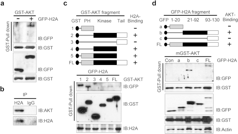
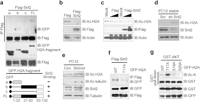
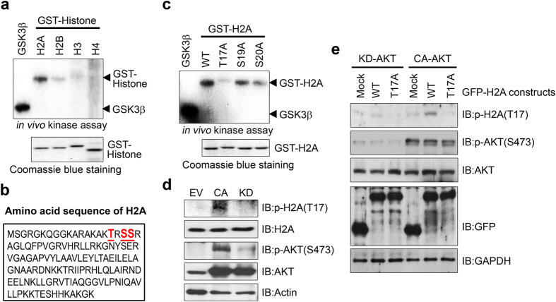
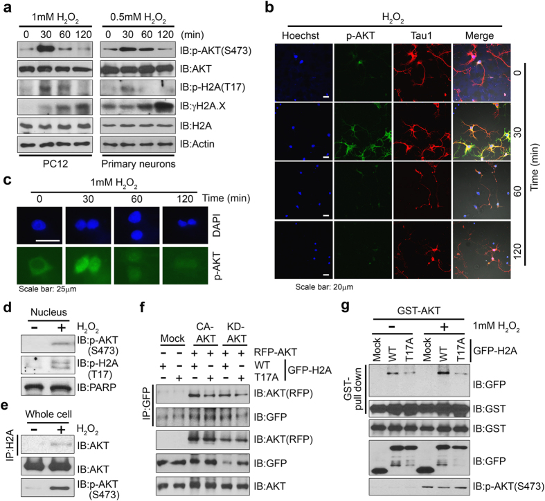
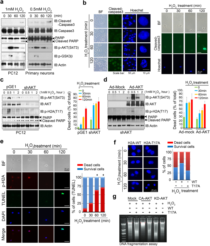
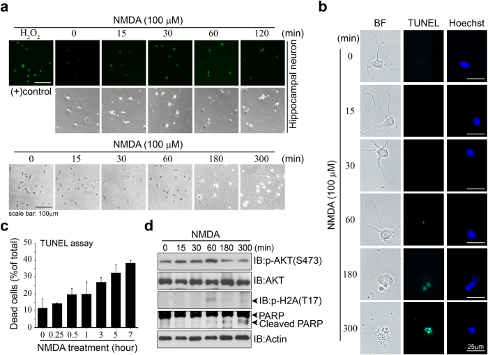
Although the essential role of protein kinase B (PKB)/Akt in cell survival signaling has been clearly established, the mechanism by which Akt mediates the cellular response to hydrogen peroxide (H2O2)-induced oxidative stress remains unclear. We demonstrated that Akt attenuated neuronal apoptosis through direct association with histone 2A (H2A) and phosphorylation of H2A at threonine 17. At early time points during H2O2 exposure of PC12 cells and primary hippocampal neurons, when the cells can tolerate the level of DNA damage, Akt was activated and phosphorylated H2A, leading to inhibition of apoptotic death. At later time points, Akt delivered the NAD(+)-dependent protein deacetylase Sirtuin 2 (Sirt 2) to the vicinity of phosphorylated H2A in response to irreversible DNA damage, thereby inducing H2A deacetylation and subsequently leading to apoptotic death. Ectopically expressed T17A-substituted H2A minimally interacted with Akt and failed to prevent apoptosis under oxidative stress. Thus Akt-mediated H2A phosphorylation has an anti-apoptotic function in conditions of H2O2-induced oxidative stress in neurons and PC12 cells.
尽管蛋白激酶B(PKB)/Akt在细胞存活信号传导中的重要作用已得到明确证实,但Akt介导细胞对过氧化氢(H2O2)诱导的氧化应激反应的机制仍不清楚。我们证明,Akt通过与组蛋白2A(H2A)直接结合并使H2A的苏氨酸17位点磷酸化来减轻神经元凋亡。在PC12细胞和原代海马神经元暴露于H2O2的早期时间点,当细胞能够耐受DNA损伤水平时,Akt被激活并使H2A磷酸化,从而抑制凋亡性死亡。在后期时间点,响应不可逆的DNA损伤,Akt将NAD(+)依赖性蛋白脱乙酰酶沉默调节蛋白2(Sirt 2)传递到磷酸化H2A附近,从而诱导H2A去乙酰化并随后导致凋亡性死亡。异位表达的T17A取代的H2A与Akt的相互作用最小,并且在氧化应激下未能预防凋亡。因此,在神经元和PC12细胞中H2O2诱导的氧化应激条件下,Akt介导的H2A磷酸化具有抗凋亡功能。