Zhu Xu, Guo Jun, He Cancan, Geng Huaxiao, Yu Gengsheng, Li Jinqing, Zheng Hairong, Ji Xiaojuan, Yan Fei
Department of Cardiology, Children's Hospital of Chongqing Medical University, Chongqing, China.
Ministry of Education Key Laboratory of Child Development and Disorders, Chongqing Key Laboratory of Pediatrics, Chongqing, China.
Sci Rep. 2016 Feb 22;6:21683. doi: 10.1038/srep21683.
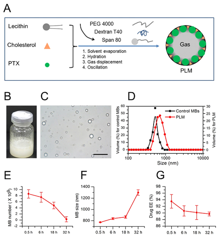
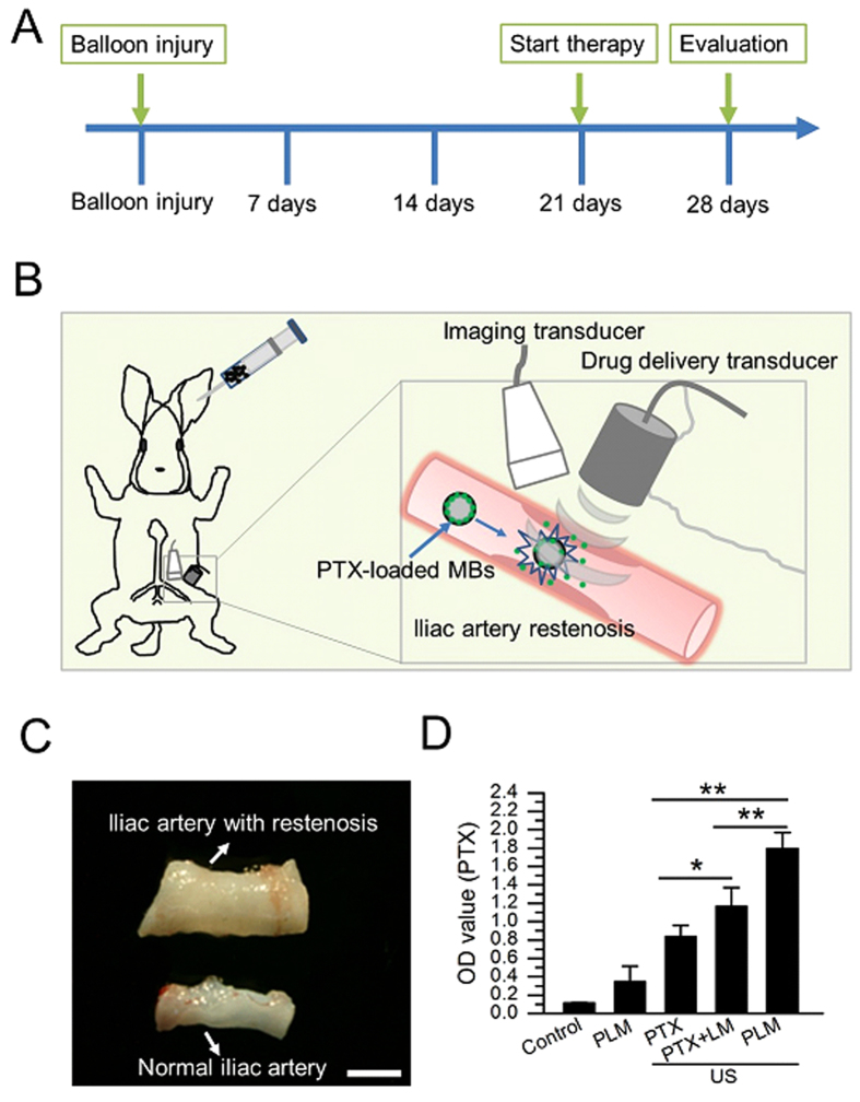
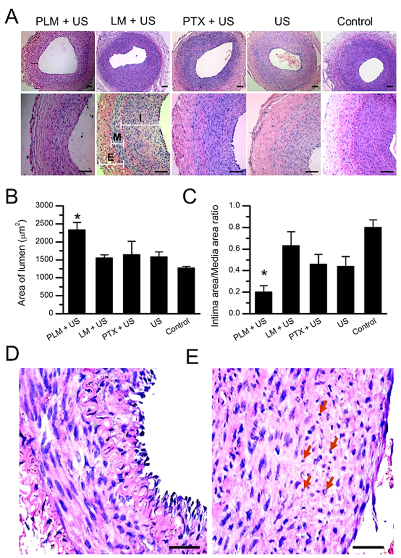
Paclitaxel (PTX) has been recognized as a promising drug for intervention of vascular reconstructions. However, it is still difficult to achieve local drug delivery in a spatio-temporally controllable manner under real-time image guidance. Here, we introduce an ultrasound (US) triggered image-guided drug delivery approach to inhibit vascular reconstruction via paclitaxel (PTX)-loaded microbubbles (PLM) in a rabbit iliac balloon injury model. PLM was prepared through encapsulating PTX in the shell of lipid microbubbles via film hydration and mechanical vibration technique. Our results showed PLM could effectively deliver PTX when exposed to US irradiation and result in significantly lower viability of vascular smooth muscle cells. Ultrasonographic examinations revealed the US signals from PLM in the iliac artery were greatly increased after intravenous administration of PLM, making it possible to identify the restenosis regions of iliac artery. The in vivo anti-restenosis experiments with PLM and US greatly inhibited neointimal hyperplasia at the injured site, showing an increased lumen area and reduced the ratio of intima area and the media area (I/M ratio). No obvious functional damages to liver and kidney were observed for those animals. Our study provided a promising approach to realize US triggered image-guided PTX delivery for therapeutic applications against iliac restenosis.
紫杉醇(PTX)已被公认为是一种用于干预血管重建的有前景的药物。然而,在实时图像引导下,以时空可控的方式实现局部药物递送仍然困难。在此,我们介绍一种超声(US)触发的图像引导药物递送方法,通过在兔髂动脉球囊损伤模型中使用负载紫杉醇(PTX)的微泡(PLM)来抑制血管重建。PLM是通过薄膜水化和机械振动技术将PTX包裹在脂质微泡的壳中制备而成。我们的结果表明,PLM在受到超声照射时能够有效地递送PTX,并导致血管平滑肌细胞的活力显著降低。超声检查显示,静脉注射PLM后,髂动脉中PLM的超声信号大大增加,从而有可能识别髂动脉的再狭窄区域。使用PLM和超声进行的体内抗再狭窄实验极大地抑制了损伤部位的内膜增生,表现为管腔面积增加,内膜面积与中膜面积之比(I/M比)降低。未观察到这些动物的肝脏和肾脏有明显的功能损害。我们的研究为实现超声触发的图像引导PTX递送用于治疗髂动脉再狭窄提供了一种有前景的方法。