Izabela Rusiecka, Jarosław Ruczyński, Magdalena Alenowicz, Piotr Rekowski, Ivan Kocić
Department of Pharmacology, Medical University of Gdańsk, Gdańsk, Poland.
Department of Chemistry, University of Gdańsk, Gdańsk, Poland.
Naunyn Schmiedebergs Arch Pharmacol. 2016 May;389(5):485-97. doi: 10.1007/s00210-016-1219-5. Epub 2016 Feb 22.
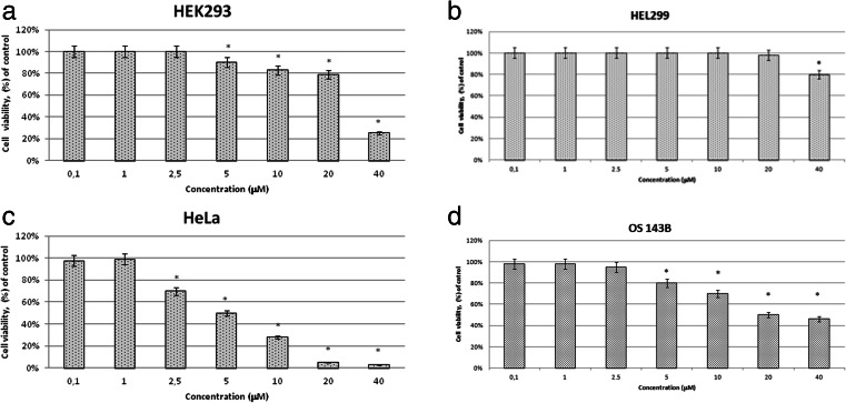
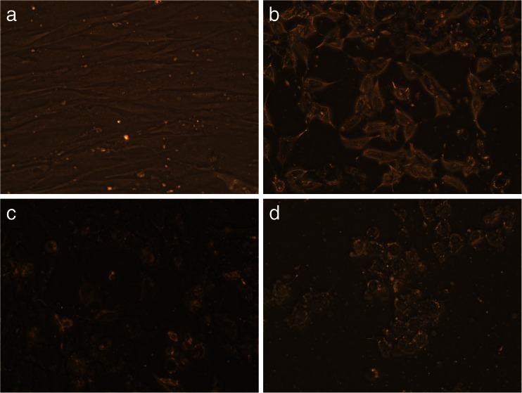
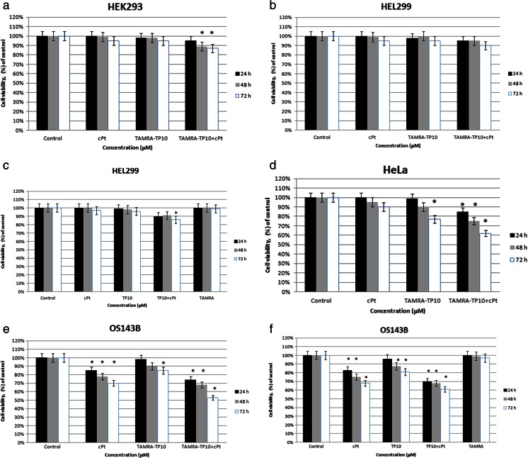
The aim of this paper was to examine whether cell-penetrating peptides (CPPs) such as transportan 10 (TP10) or protein transduction domain (PTD4) may improve the anticancer activity of cisplatin (cPt). The complexes of TP10 or PTD4 with cPt were used in the experiments. They were carried out on two non-cancer (HEK293 (human embryonic kidney) and HEL299 (human embryo lung)) and two cancer (HeLa (human cervical cancer) and OS143B (human osteosarcoma 143B)) cell lines. Both complexes were tested (MTT assay) with respect to their anticancer or cytotoxic actions. TAMRA (fluorescent dye)-stained preparations were visualized in a fluorescence microscope. The long-term effect of TP10 + cPt and its components on non-cancer and cancer cell lines was observed in inverted phase contrast microscopy. In the MTT test (cell viability assay), the complex of TP10 + cPt produced a more potent effect on the cancer cell lines (HeLa, OS143B) in comparison to that observed after separate treatment with TP10 or cPt. At the same time, the action of the complex and its components was rather small on non-cancer cell lines. On the other hand, a complex of another CPP with cPt, i.e., PTD4 + cPt, was without a significant effect on the cancer cell line (OS143B). The images of the fluorescent microscopy showed TAMRA-TP10 or TAMRA-TP10 + cPt in the interior of the HeLa cells. In the case of TAMRA-PTD4 or TAMRA-PTD4 + cPt, only the first compound was found inside the cancer cell line. In contrast, none of the tested compounds gained access to the interior of the non-cancer cells (HEK293, HEL299). Long-term incubation with the TP10 + cPt (estimated by inverted phase contrast microscopy) lead to an enhanced action of the complex on cell viability (decrease in the number of cells and change in their morphology) as compared with that produced by each single agent. With regard to the tested CPPs, only TP10 improved the anticancer activity of cisplatin if both compounds were used in the form of a complex. Additionally, the complex was relatively safe for non-cancer cells. What is more, TP10 also produced an anticancer effect on HeLa and OS143B cell lines.
本文的目的是研究穿膜肽,如运输蛋白10(TP10)或蛋白转导结构域(PTD4),是否可以提高顺铂(cPt)的抗癌活性。实验中使用了TP10或PTD4与cPt的复合物。实验在两种非癌细胞系(HEK293(人胚肾)和HEL299(人胚肺))以及两种癌细胞系(HeLa(人宫颈癌)和OS143B(人骨肉瘤143B))上进行。通过MTT法检测了这两种复合物的抗癌或细胞毒性作用。用荧光显微镜观察了四甲基罗丹明(TAMRA,荧光染料)染色的制剂。通过倒置相差显微镜观察了TP10 + cPt及其成分对非癌细胞系和癌细胞系的长期影响。在MTT试验(细胞活力测定)中,与单独使用TP10或cPt相比,TP10 + cPt复合物对癌细胞系(HeLa、OS143B)产生了更强的作用。同时,该复合物及其成分对非癌细胞系的作用较小。另一方面,另一种CPP与cPt的复合物,即PTD4 + cPt,对癌细胞系(OS143B)没有显著影响。荧光显微镜图像显示HeLa细胞内部有TAMRA-TP10或TAMRA-TP10 + cPt。对于TAMRA-PTD4或TAMRA-PTD4 + cPt,仅在癌细胞系中发现了第一种化合物。相反,所测试的化合物均未进入非癌细胞(HEK293、HEL299)内部。与每种单一药物相比,通过倒置相差显微镜估计,用TP10 + cPt长期孵育导致该复合物对细胞活力的作用增强(细胞数量减少及其形态改变)。关于所测试的穿膜肽,如果两种化合物以复合物形式使用,只有TP10提高了顺铂的抗癌活性。此外,该复合物对非癌细胞相对安全。而且,TP10对HeLa和OS143B细胞系也产生了抗癌作用。