Sprecher Kate E, Riedner Brady A, Smith Richard F, Tononi Giulio, Davidson Richard J, Benca Ruth M
Department of Psychiatry, University of Wisconsin, Madison, Wisconsin, United States of America.
Wisconsin Center for Sleep Medicine and Research, University of Wisconsin, Madison, Wisconsin, United States of America.
PLoS One. 2016 Feb 22;11(2):e0149770. doi: 10.1371/journal.pone.0149770. eCollection 2016.
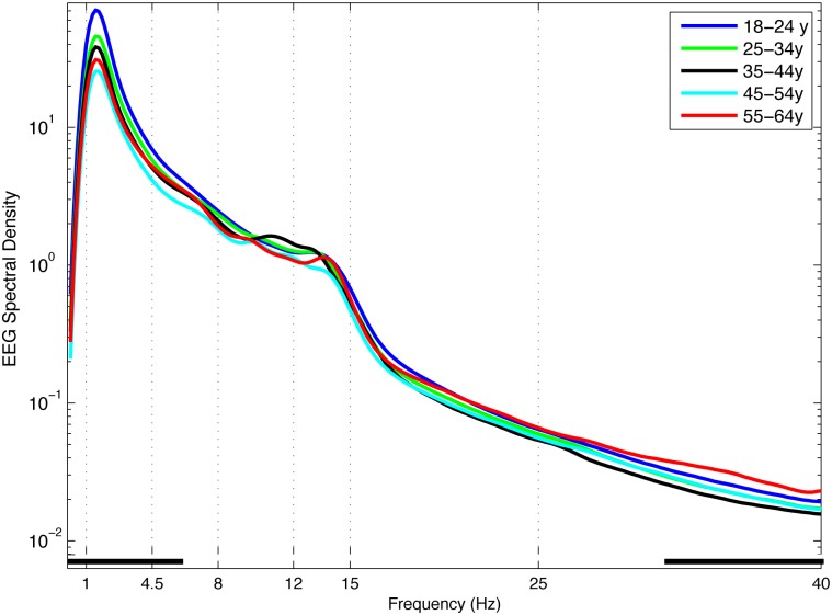
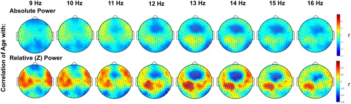
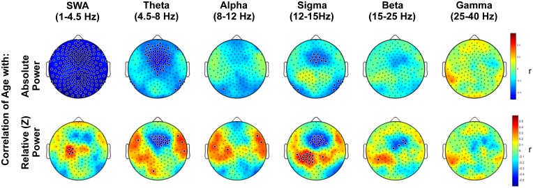
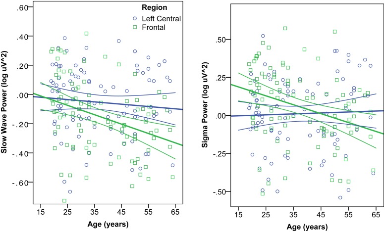
Sleeping brain activity reflects brain anatomy and physiology. The aim of this study was to use high density (256 channel) electroencephalography (EEG) during sleep to characterize topographic changes in sleep EEG power across normal aging, with high spatial resolution. Sleep was evaluated in 92 healthy adults aged 18-65 years old using full polysomnography and high density EEG. After artifact removal, spectral power density was calculated for standard frequency bands for all channels, averaged across the NREM periods of the first 3 sleep cycles. To quantify topographic changes with age, maps were generated of the Pearson's coefficient of the correlation between power and age at each electrode. Significant correlations were determined by statistical non-parametric mapping. Absolute slow wave power declined significantly with increasing age across the entire scalp, whereas declines in theta and sigma power were significant only in frontal regions. Power in fast spindle frequencies declined significantly with increasing age frontally, whereas absolute power of slow spindle frequencies showed no significant change with age. When EEG power was normalized across the scalp, a left centro-parietal region showed significantly less age-related decline in power than the rest of the scalp. This partial preservation was particularly significant in the slow wave and sigma bands. The effect of age on sleep EEG varies substantially by region and frequency band. This non-uniformity should inform the design of future investigations of aging and sleep. This study provides normative data on the effect of age on sleep EEG topography, and provides a basis from which to explore the mechanisms of normal aging as well as neurodegenerative disorders for which age is a risk factor.
睡眠脑电活动反映大脑的解剖结构和生理机能。本研究旨在利用睡眠期间的高密度(256导)脑电图(EEG),以高空间分辨率表征正常衰老过程中睡眠EEG功率的地形学变化。使用全夜多导睡眠图和高密度EEG对92名年龄在18至65岁的健康成年人的睡眠进行评估。去除伪迹后,计算所有通道标准频段的频谱功率密度,并在前3个睡眠周期的非快速眼动期进行平均。为了量化随年龄的地形学变化,生成了每个电极处功率与年龄之间皮尔逊相关系数的图谱。通过统计非参数映射确定显著相关性。在整个头皮上,绝对慢波功率随年龄增长显著下降,而θ波和σ波功率仅在额叶区域显著下降。快速纺锤波频率的功率在额叶随年龄增长显著下降,而慢速纺锤波频率的绝对功率随年龄无显著变化。当头皮上的EEG功率进行标准化时,左侧中央顶叶区域的功率与年龄相关的下降明显小于头皮其他部位。这种部分保留在慢波和σ波段尤为显著。年龄对睡眠EEG的影响因区域和频段而异。这种不均匀性应为未来衰老与睡眠研究的设计提供参考。本研究提供了年龄对睡眠EEG地形学影响的规范性数据,并为探索正常衰老机制以及年龄作为危险因素的神经退行性疾病机制提供了基础。