Fitzroy Ahren B, Jones Bethany J, Kainec Kyle A, Seo Jeehye, Spencer Rebecca M C
Neuroscience & Behavior Program, University of Massachusetts Amherst, Amherst, MA, United States.
Department of Psychological and Brain Sciences, University of Massachusetts Amherst, Amherst, MA, United States.
Front Aging Neurosci. 2022 Jan 11;13:787654. doi: 10.3389/fnagi.2021.787654. eCollection 2021.
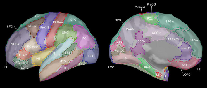
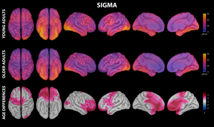
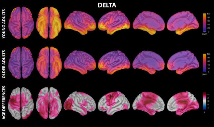
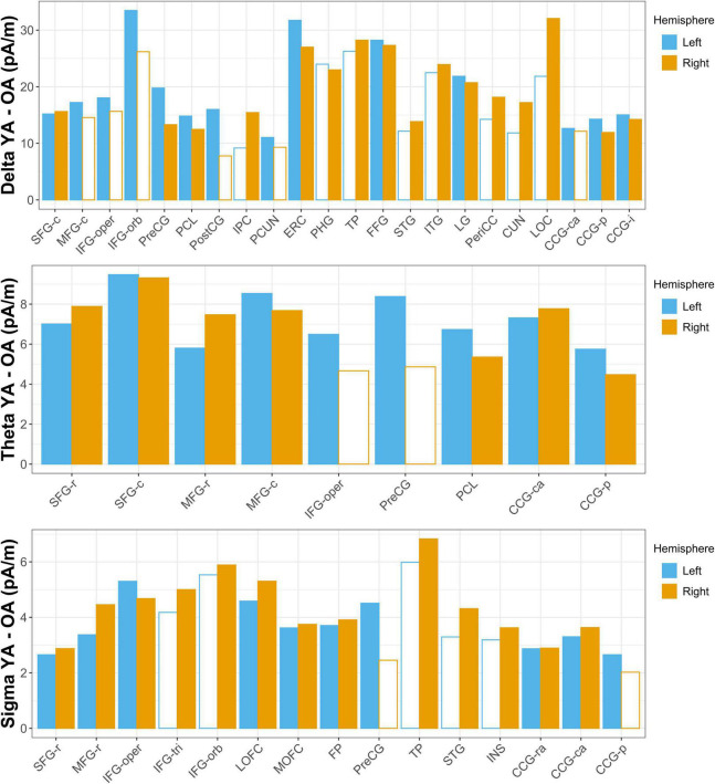
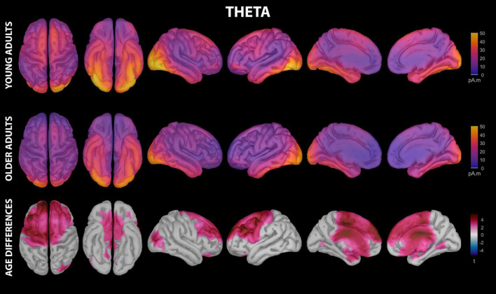
Oscillatory neural activity during sleep, such as that in the delta and sigma bands, is important for motor learning consolidation. This activity is reduced with typical aging, and this reduction may contribute to aging-related declines in motor learning consolidation. Evidence suggests that brain regions involved in motor learning contribute to oscillatory neural activity during subsequent sleep. However, aging-related differences in regional contributions to sleep oscillatory activity following motor learning are unclear. To characterize these differences, we estimated the cortical sources of consolidation-related oscillatory activity using individual anatomical information in young and older adults during non-rapid eye movement sleep after motor learning and analyzed them in light of cortical thickness and pre-sleep functional brain activation. High-density electroencephalogram was recorded from young and older adults during a midday nap, following completion of a functional magnetic resonance imaged serial reaction time task as part of a larger experimental protocol. Sleep delta activity was reduced with age in a left-weighted motor cortical network, including premotor cortex, primary motor cortex, supplementary motor area, and pre-supplementary motor area, as well as non-motor regions in parietal, temporal, occipital, and cingulate cortices. Sleep theta activity was reduced with age in a similar left-weighted motor network, and in non-motor prefrontal and middle cingulate regions. Sleep sigma activity was reduced with age in left primary motor cortex, in a non-motor right-weighted prefrontal-temporal network, and in cingulate regions. Cortical thinning mediated aging-related sigma reductions in lateral orbitofrontal cortex and frontal pole, and partially mediated delta reductions in parahippocampal, fusiform, and lingual gyri. Putamen, caudate, and inferior parietal cortex activation prior to sleep predicted frontal and motor cortical contributions to sleep delta and theta activity in an age-moderated fashion, reflecting negative relationships in young adults and positive or absent relationships in older adults. Overall, these results support the local sleep hypothesis that brain regions active during learning contribute to consolidation-related neural activity during subsequent sleep and demonstrate that sleep oscillatory activity in these regions is reduced with aging.
睡眠期间的振荡性神经活动,如δ波和σ波频段的活动,对运动学习巩固很重要。这种活动会随着典型衰老而减少,而这种减少可能导致与衰老相关的运动学习巩固能力下降。有证据表明,参与运动学习的脑区会在随后的睡眠中对振荡性神经活动产生影响。然而,运动学习后区域对睡眠振荡活动的贡献在衰老过程中的差异尚不清楚。为了描述这些差异,我们利用年轻和年长成年人在运动学习后的非快速眼动睡眠期间的个体解剖信息,估计了与巩固相关的振荡活动的皮质源,并根据皮质厚度和睡眠前功能性脑激活情况对其进行了分析。在一项功能磁共振成像序列反应时任务完成后,于午间小睡期间记录了年轻和年长成年人的高密度脑电图,该任务是一个更大实验方案的一部分。在包括运动前皮质、初级运动皮质、辅助运动区和前辅助运动区在内的左侧加权运动皮质网络以及顶叶、颞叶、枕叶和扣带回皮质的非运动区域,睡眠δ波活动随年龄增长而减少。在类似的左侧加权运动网络以及非运动前额叶和中扣带回区域,睡眠θ波活动随年龄增长而减少。在左侧初级运动皮质、右侧加权的非运动前额叶 - 颞叶网络以及扣带回区域,睡眠σ波活动随年龄增长而减少。皮质变薄介导了外侧眶额皮质和额极与衰老相关的σ波减少,并部分介导了海马旁回、梭状回和舌回的δ波减少。睡眠前壳核、尾状核和顶下小叶皮质的激活以年龄调节的方式预测了额叶和运动皮质对睡眠δ波和θ波活动的贡献,反映出年轻成年人中的负相关关系以及年长成年人中的正相关关系或无相关关系。总体而言,这些结果支持局部睡眠假说,即学习期间活跃的脑区会对随后睡眠中与巩固相关的神经活动产生影响,并表明这些区域的睡眠振荡活动会随着衰老而减少。