Pahuja Natasha Kishore, Shetty Rohit, Nuijts Rudy M M A, Agrawal Aarti, Ghosh Arkasubhra, Jayadev Chaitra, Nagaraja Harsha
Narayana Nethralaya Eye Hospital, Bangalore 560010, India.
Academic Hospital, Maastricht University, P.O. Box 616, 6200 MD Maastricht, Netherlands.
Biomed Res Int. 2016;2016:5067853. doi: 10.1155/2016/5067853. Epub 2016 Jan 21.
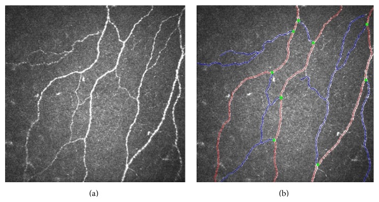
Purpose. To study the corneal nerve morphology and its importance in unilateral keratoconus. Materials and Methods. In this prospective cross-sectional study, 33 eyes of 33 patients with keratoconus in one eye (Group 3) were compared with the other normal eye of the same patients (Group 2) and 30 eyes of healthy patients (Group 1). All patients underwent detailed ophthalmic examination followed by topography with Pentacam HR and in vivo confocal microscopy (IVCM). Five images obtained with IVCM were analyzed using an automated CCmetrics software version 1.0 for changes in subbasal plexus of nerves. Results. Intergroup comparison showed statistically significant reduction in corneal nerve fiber density (CNFD) and length (CNFL) in Group 3 as compared to Group 1 (p < 0.001 and p = 0.001, resp.) and Group 2 (p = 0.01 and p = 0.02, resp.). Though corneal nerve fiber length, diameter, area, width, corneal nerve branch density, and corneal total branch density were found to be higher in decentered cones, only the corneal nerve branch density (CNBD) was found to be statistically significant (p < 0.01) as compared to centered cones. Conclusion. Quantitative changes in the corneal nerve morphology can be used as an imaging marker for the early diagnosis of keratoconus before the onset of refractive or topography changes.
目的。研究单侧圆锥角膜的角膜神经形态及其重要性。材料与方法。在这项前瞻性横断面研究中,将33例单眼圆锥角膜患者的33只患眼(第3组)与同一患者的另一只正常眼(第2组)以及30例健康患者的30只眼(第1组)进行比较。所有患者均接受详细的眼科检查,随后使用Pentacam HR进行地形图检查以及活体共聚焦显微镜检查(IVCM)。使用自动化的CCmetrics软件版本1.0分析通过IVCM获得的五张图像,以观察神经基底丛的变化。结果。组间比较显示,与第1组(分别为p < 0.001和p = 0.001)和第2组(分别为p = 0.01和p = 0.02)相比,第3组的角膜神经纤维密度(CNFD)和长度(CNFL)在统计学上有显著降低。尽管发现偏心圆锥角膜的角膜神经纤维长度、直径、面积、宽度、角膜神经分支密度和角膜总分支密度更高,但与中心圆锥角膜相比,仅角膜神经分支密度(CNBD)在统计学上有显著差异(p < 0.01)。结论。角膜神经形态的定量变化可作为圆锥角膜在屈光或地形图改变出现之前早期诊断的影像学标志物。