Yamaguchi Takefumi, Hamrah Pedram, Shimazaki Jun
*Department of Ophthalmology, Ichikawa General Hospital, Tokyo Dental College, Chiba, Japan; and †Center for Translational Ocular Immunology, Department of Ophthalmology, Tufts Medical Center, Tufts University School of Medicine, Boston, MA.
Cornea. 2016 Nov;35 Suppl 1(Suppl 1):S65-S70. doi: 10.1097/ICO.0000000000000989.
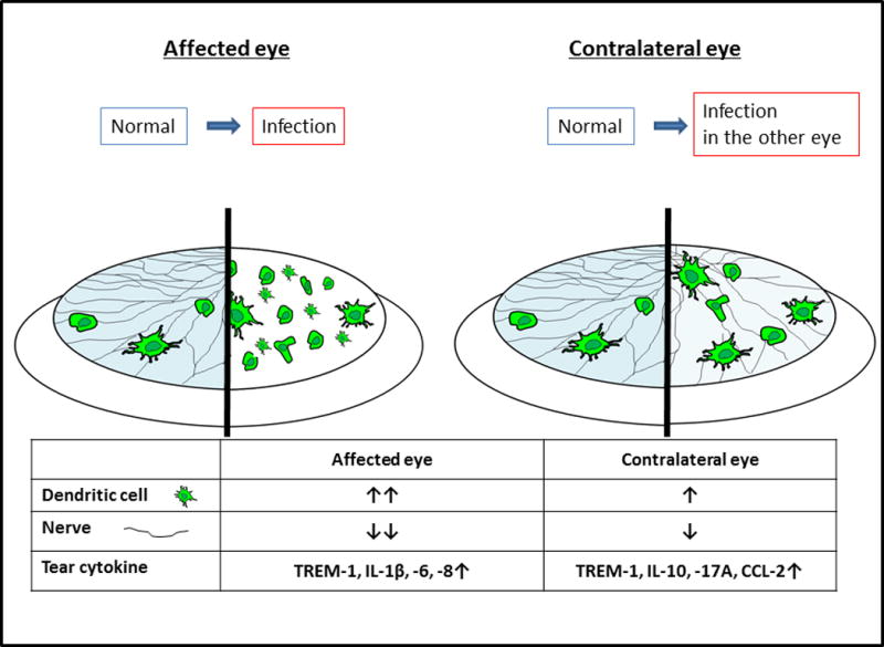
This review summarizes the recent literature regarding corneal imaging in human subjects using in vivo confocal microscopy. It also covers the recent literature on corneal immune cells, nerves, and tear cytokine levels in ocular surface diseases as well as corneal immune privilege. The significance of interactions between corneal immune cells and nerves in health, neurotrophic keratopathy, and infectious keratitis is discussed. Furthermore, bilateral alterations of immune cells and nerves in clinically unilateral corneal diseases and the link to changes of tear cytokines or neuropeptide levels in contralateral eyes are described. Recent studies reported increased density and morphologic changes of corneal dendritic cells in ocular surface disease that correlated with a decrease in subbasal nerve and corneal nerve density, suggesting potential interactions between the immune and nervous systems in the cornea. Although the relevance of tear cytokines is poorly understood, tear cytokines might have an important role in the pathogenesis of ocular surface diseases. In humans and experimental animal models, alterations in immune cells, cytokines, and immunomodulatory neuropeptide levels in contralateral eyes might mediate the incidence of bilateral infectious keratitis and loss of immune privilege of the cornea in bilateral corneal transplantation or neurotrophic keratopathy cases. The discovery of bilateral alterations of immune cells and nerves in ocular surface diseases is considered the missing link between the immune and nervous systems in the cornea, and demonstrates how studies of animal models and humans aid our understanding of human corneal disease phenomena.
本综述总结了近期有关使用活体共聚焦显微镜对人类受试者进行角膜成像的文献。它还涵盖了近期关于眼表疾病中角膜免疫细胞、神经和泪液细胞因子水平以及角膜免疫赦免的文献。讨论了角膜免疫细胞与神经在健康状态、神经营养性角膜病变和感染性角膜炎中的相互作用的意义。此外,还描述了临床单侧角膜疾病中免疫细胞和神经的双侧改变以及与对侧眼泪液细胞因子或神经肽水平变化的联系。近期研究报道,眼表疾病中角膜树突状细胞密度增加和形态改变,这与基底膜下神经和角膜神经密度降低相关,提示角膜中免疫和神经系统之间可能存在相互作用。尽管对泪液细胞因子的相关性了解甚少,但泪液细胞因子可能在眼表疾病的发病机制中起重要作用。在人类和实验动物模型中,对侧眼角膜免疫细胞、细胞因子和免疫调节神经肽水平的改变可能介导双侧感染性角膜炎的发生以及双侧角膜移植或神经营养性角膜病变病例中角膜免疫赦免的丧失。眼表疾病中免疫细胞和神经的双侧改变的发现被认为是角膜免疫和神经系统之间缺失的环节,并展示了动物模型和人类研究如何帮助我们理解人类角膜疾病现象。