Brunner Gerd, Bismuth Jean, Nambi Vijay, Ballantyne Christie M, Taylor Addison A, Lumsden Alan B, Morrisett Joel D, Shah Dipan J
Section of Cardiovascular Research, Department of Medicine, Baylor College of Medicine, Houston, TX, USA.
Methodist DeBakey Heart and Vascular Center, Houston Methodist Hospital, 6565 Fannin St., FB-Alkek Tower - Suite A679A, Mailstop A-601, Houston, TX, 77030, USA.
Med Biol Eng Comput. 2016 Nov;54(11):1667-1681. doi: 10.1007/s11517-016-1457-1. Epub 2016 Feb 23.
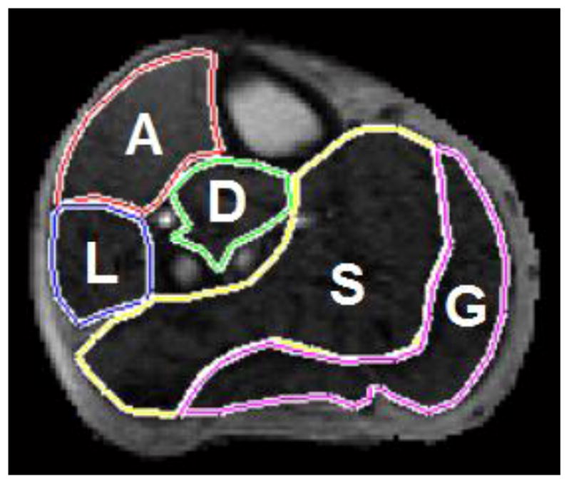
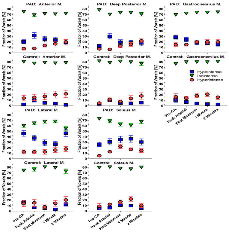
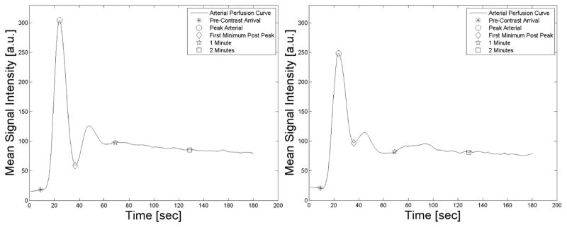
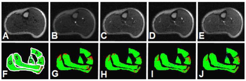
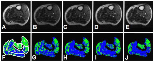
We hypothesized that skeletal muscle perfusion is impaired in peripheral arterial disease (PAD) patients compared to healthy controls and that perfusion patterns exhibit marked differences across five leg muscle compartments including the anterior muscle group (AM), lateral muscle group (LM), deep posterior muscle group (DM), soleus (SM), and the gastrocnemius muscle (GM). A total of 40 individuals (26 PAD patients and 14 healthy controls) underwent contrast-enhanced magnetic resonance imaging (CE-MRI) utilizing a reactive hyperemia protocol. Muscle perfusion maps were developed for AM, LM, DM, SM, and GM. Perfusion maps were analyzed over the course of 2 min, starting at local pre-contrast arrival, to study early-to-intermediate gadolinium enhancement. PAD patients had a higher fraction of hypointense voxels at pre-contrast arrival for all five muscle compartments compared with healthy controls (p < 0.0005). Among PAD patients, the fraction of hypointense voxels of the AM, LM, and GM were inversely correlated with the estimated glomerular filtration rate (eGFR; r = -0.509, p = 0.008; r = -0.441, p = 0.024; and r = -0.431, p = 0.028, respectively). CE-MRI-based skeletal leg muscle perfusion is markedly reduced in PAD patients compared with healthy controls and shows heterogeneous patterns across calf muscle compartments.
我们假设,与健康对照组相比,外周动脉疾病(PAD)患者的骨骼肌灌注受损,并且灌注模式在五个腿部肌肉区之间存在显著差异,这五个肌肉区包括前肌群(AM)、外侧肌群(LM)、后深部肌群(DM)、比目鱼肌(SM)和腓肠肌(GM)。共有40名个体(26名PAD患者和14名健康对照者)采用反应性充血方案接受了对比增强磁共振成像(CE-MRI)检查。为AM、LM、DM、SM和GM绘制了肌肉灌注图。从局部对比剂到达前开始,在2分钟的过程中分析灌注图,以研究钆剂的早期至中期增强情况。与健康对照组相比,PAD患者在对比剂到达前,所有五个肌肉区的低信号体素比例更高(p < 0.0005)。在PAD患者中,AM、LM和GM的低信号体素比例与估计肾小球滤过率(eGFR)呈负相关(分别为r = -0.509,p = 0.008;r = -0.441,p = 0.024;r = -0.431,p = 0.028)。与健康对照组相比,基于CE-MRI的PAD患者腿部骨骼肌灌注明显降低,并且在小腿肌肉区呈现出异质性模式。