Cardiovascular Imaging Research and Data Sciences Laboratory, Department of Medicine, Baylor College of Medicine, Houston, TX, USA; Penn State Heart and Vascular Institute, Pennsylvania State University College of Medicine, Hershey, PA, USA.
Methodist DeBakey Heart and Vascular Center, Houston Methodist Hospital, Houston, TX, USA.
J Biomech. 2019 Aug 27;93:147-158. doi: 10.1016/j.jbiomech.2019.06.025. Epub 2019 Jul 8.
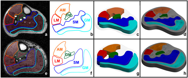
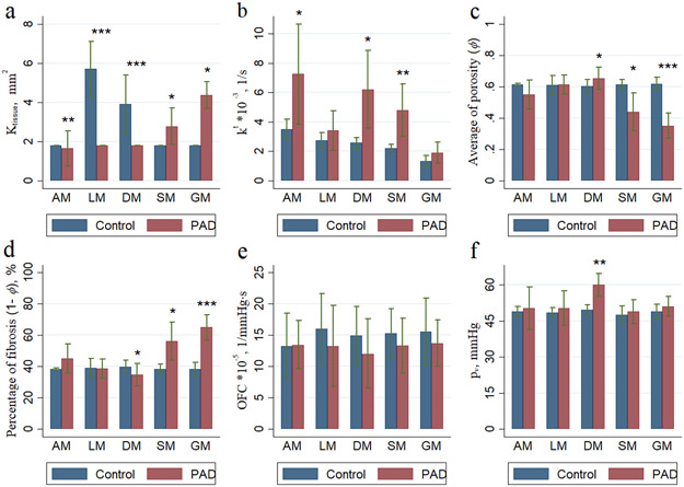
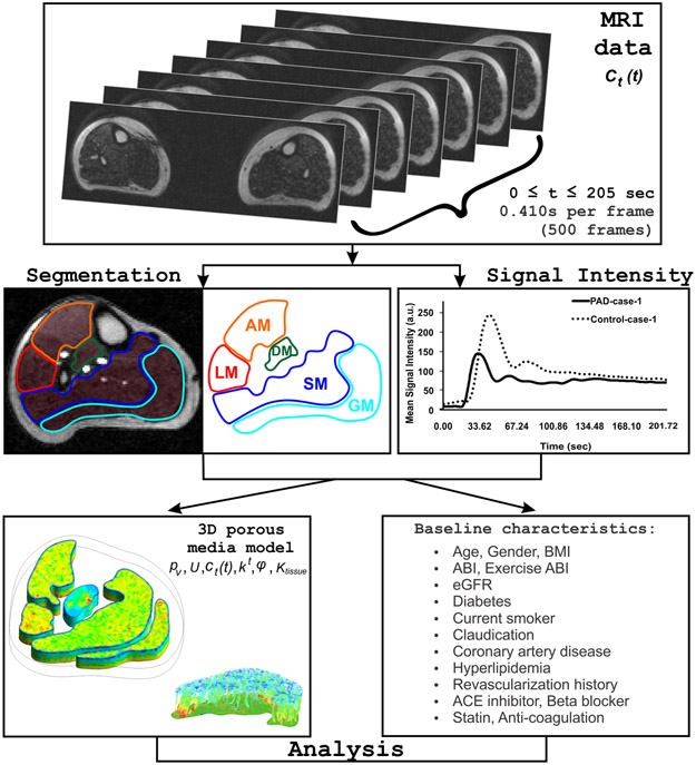
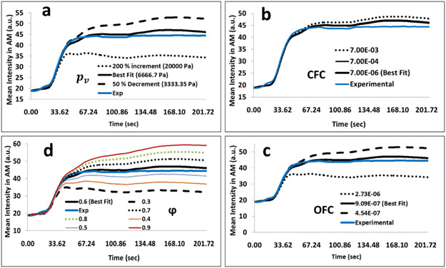
Peripheral artery disease (PAD) is associated with an increased risk of adverse cardiovascular events, impaired lower extremity blood flow and microvascular perfusion abnormalities in the calf muscles which can be determined with contrast-enhanced magnetic resonance imaging (CE-MRI). We developed a computational model of the microvascular perfusion in the calf muscles. We included 20 patients (10 PAD, 10 controls) and utilized the geometry, mean signal intensity and arterial input functions from CE-MRI calf muscle perfusion scans. The model included the microvascular pressure (p), outflow filtration coefficient (OFC), transfer rate constant (k), porosity (φ), and the interstitial permeability (K). Parameters were fitted and the simulations were compared across PAD patients and controls. Intra-observer reproducibility of the simulated mean signal intensities was excellent (intraclass correlation coefficients >0.995). k and K were higher in PAD patients compared with controls (4.72 interquartile range (IQR) 3.33, 5.56 vs. 2.47 IQR 2.10, 2.85; p = 0.003; and 3.68 IQR 3.18, 4.41 vs. 1.81 IQR 1.81, 1.81; p < 0.001). Conversely, porosity (φ) was lower in PAD patients compared with controls (0.52 IQR 0.49, 0.54 vs. 0.61 IQR 0.58, 0.64; p = 0.016). Porosity (φ) was correlated with the ankle brachial index (r = 0.64, p = 0.011). The proposed computational microvascular model is robust and reproducible, and essential model parameters differ significantly between PAD patients and controls.
外周动脉疾病(PAD)与不良心血管事件风险增加、下肢血流受损和小腿肌肉微血管灌注异常有关,这些可以通过对比增强磁共振成像(CE-MRI)来确定。我们开发了一种小腿肌肉微血管灌注的计算模型。我们纳入了 20 名患者(10 名 PAD 患者,10 名对照者),并利用 CE-MRI 小腿肌肉灌注扫描的几何形状、平均信号强度和动脉输入函数。该模型包括微血管压力(p)、流出过滤系数(OFC)、转运率常数(k)、孔隙率(φ)和间质通透性(K)。对参数进行拟合,并比较 PAD 患者和对照组的模拟平均信号强度。模拟平均信号强度的观察者内可重复性非常好(组内相关系数>0.995)。与对照组相比,PAD 患者的 k 和 K 更高(4.72 四分位间距(IQR)3.33、5.56 与 2.47 IQR 2.10、2.85;p=0.003;3.68 IQR 3.18、4.41 与 1.81 IQR 1.81、1.81;p<0.001)。相反,与对照组相比,PAD 患者的孔隙率(φ)更低(0.52 IQR 0.49、0.54 与 0.61 IQR 0.58、0.64;p=0.016)。孔隙率(φ)与踝臂指数(ABI)相关(r=0.64,p=0.011)。提出的计算微血管模型具有稳健性和可重复性,并且 PAD 患者和对照组之间的基本模型参数有显著差异。