Broersma Marja, van der Stouwe Anna M M, Buijink Arthur W G, de Jong Bauke M, Groot Paul F C, Speelman Johannes D, Tijssen Marina A J, van Rootselaar Anne-Fleur, Maurits Natasha M
Department of Neurology, University Medical Center Groningen, University of Groningen, PO Box 30001, 9700 RB Groningen, the Netherlands; Neuroimaging Center, University Medical Center Groningen, University of Groningen, PO Box 30001, 9700 RB Groningen, the Netherlands.
Department of Neurology and Clinical Neurophysiology, Academic Medical Center, University of Amsterdam, PO Box 22660, 1100 DD Amsterdam, the Netherlands; Brain Imaging Center, Academic Medical Center, University of Amsterdam, PO Box 22660, 1100 DD Amsterdam, the Netherlands.
Neuroimage Clin. 2015 Dec 28;11:1-9. doi: 10.1016/j.nicl.2015.12.011. eCollection 2016.
Essential tremor (ET) is one of the most common hyperkinetic movement disorders. Previous research into the pathophysiology of ET suggested underlying cerebellar abnormalities.
In this study, we added electromyography as an index of tremor intensity to functional Magnetic Resonance Imaging (EMG-fMRI) to study a group of ET patients selected according to strict criteria to achieve maximal homogeneity. With this approach we expected to improve upon the localization of the bilateral cerebellar abnormalities found in earlier fMRI studies.
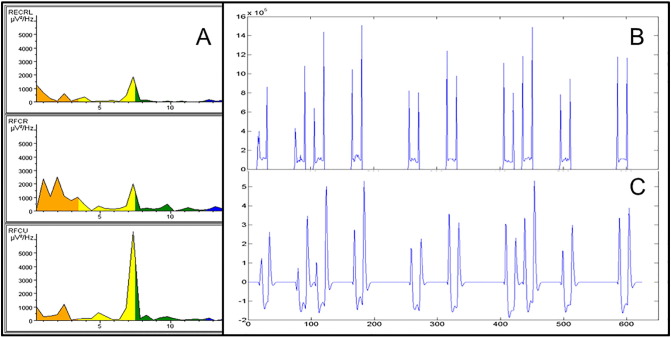
We included 21 propranolol sensitive patients, who were not using other tremor medication, with a definite diagnosis of ET defined by the Tremor Investigation Group. Simultaneous EMG-fMRI recordings were performed while patients were off tremor medication. Patients performed unilateral right hand and arm extension, inducing tremor, alternated with relaxation (rest). Twenty-one healthy, age- and sex-matched participants mimicked tremor during right arm extension. EMG power variability at the individual tremor frequency as a measure of tremor intensity variability was used as a regressor, mathematically independent of the block regressor, in the general linear model used for fMRI analysis, to find specific tremor-related activations.
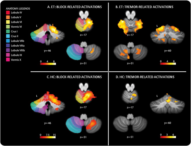
Block-related activations were found in the classical upper-limb motor network, both for ET patients and healthy participants in motor, premotor and supplementary motor areas. In ET patients, we found tremor-related activations bilaterally in the cerebellum: in left lobules V, VI, VIIb and IX and in right lobules V, VI, VIIIa and b, and in the brainstem. In healthy controls we found simulated tremor-related activations in right cerebellar lobule V.
Our results expand on previous findings of bilateral cerebellar involvement in ET. We have identified specific areas in the bilateral somatomotor regions of the cerebellum: lobules V, VI and VIII.
特发性震颤(ET)是最常见的运动亢进性运动障碍之一。先前对ET病理生理学的研究表明存在潜在的小脑异常。
在本研究中,我们将肌电图作为震颤强度指标添加到功能磁共振成像(EMG-fMRI)中,以研究一组根据严格标准选择的ET患者,以实现最大同质性。通过这种方法,我们期望改进早期fMRI研究中发现的双侧小脑异常的定位。
我们纳入了21名对普萘洛尔敏感且未使用其他震颤药物的患者,这些患者由震颤研究小组明确诊断为ET。在患者停用震颤药物时进行同步EMG-fMRI记录。患者进行右手和右臂单侧伸展,诱发震颤,与放松(休息)交替进行。21名年龄和性别匹配的健康参与者在右臂伸展时模拟震颤。在用于fMRI分析的一般线性模型中,将个体震颤频率下的EMG功率变异性作为震颤强度变异性的度量用作回归变量,在数学上独立于块回归变量,以找到特定的震颤相关激活。
在经典的上肢运动网络中发现了与块相关的激活,ET患者和健康参与者在运动、运动前区和辅助运动区均有激活。在ET患者中,我们在小脑双侧发现了与震颤相关的激活:在左侧小叶V、VI、VIIb和IX以及右侧小叶V、VI、VIIIa和b以及脑干中。在健康对照中,我们在右侧小脑小叶V中发现了模拟的震颤相关激活。
我们的结果扩展了先前关于ET双侧小脑受累的发现。我们已经确定了小脑双侧躯体运动区域中的特定区域:小叶V、VI和VIII。