Koroglu Reyhan, Koksal Ismail, Gezer Fatma, Kahraman Aysegul, Kekilli Ersoy
Department of Nuclear Medicine, Karabuk University, Karabuk, Turkey.
Department of Nuclear Medicine, Inonu University, Turgut Ozal Medical Center, Malatya, Turkey.
World J Nucl Med. 2016 Jan-Apr;15(1):68-70. doi: 10.4103/1450-1147.167590.
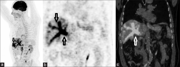
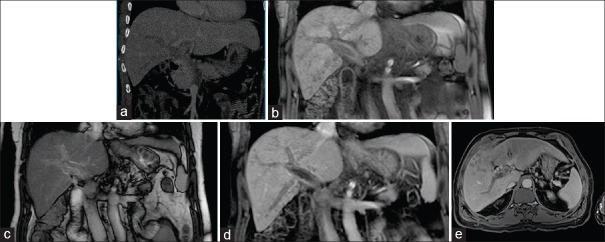
Major vascular invasion is one of the worst prognostic factors of hepatocellular carcinoma (HCC). Fludeoxyglucose F 18 ((18) F-FDG) positron emission tomography/computed tomography (PET/CT) method is succesfully being used in HCC patients for the detection of particularly long-distance metastasis. Major vascular invasion is shown by radiological methods [particularly dynamic CT and/or magnetic resonance imaging (MRI)]. A male patient aged 60 years was diagnosed with HCC, according to biopsy after the detection of a mass in the liver. His medical examinations that were performed for the evaluation in terms of liver transplantation were dynamic CT and dynamic MRI; invasion in the intrahepatic branches of the portal vein and in main portal vein was also detected. PET/CT was performed to investigate the distant metastases. Moreover, diffuse (18) F-FDG uptake in the intrahepatic branches of the portal vein and in the main portal vein was observed.
大血管侵犯是肝细胞癌(HCC)最糟糕的预后因素之一。氟脱氧葡萄糖F 18((18)F-FDG)正电子发射断层扫描/计算机断层扫描(PET/CT)方法已成功应用于HCC患者,用于检测特别是远距离转移。大血管侵犯通过放射学方法(特别是动态CT和/或磁共振成像(MRI))显示。一名60岁男性患者在肝脏发现肿块后经活检确诊为HCC。为评估肝移植进行的医学检查是动态CT和动态MRI;还检测到门静脉肝内分支和主门静脉有侵犯。进行PET/CT以调查远处转移。此外,观察到门静脉肝内分支和主门静脉有弥漫性(18)F-FDG摄取。