He Yanyu, Yu Sijiu, Hu Junwei, Cui Yan, Liu Penggang
College of Veterinary Medicine, Gansu Agricultural University, Lanzhou 730070, Gansu, China.
PLoS One. 2016 Feb 25;11(2):e0149947. doi: 10.1371/journal.pone.0149947. eCollection 2016.
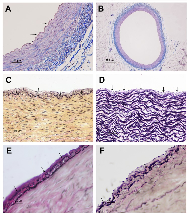
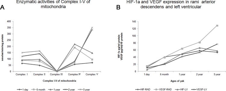
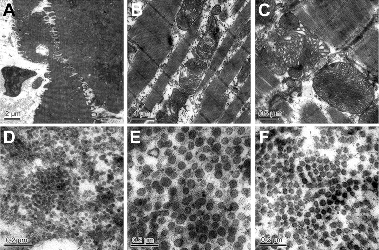
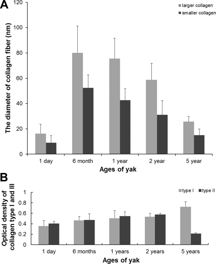
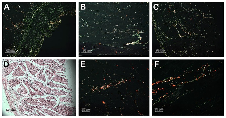
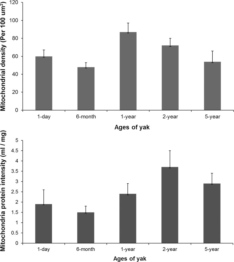
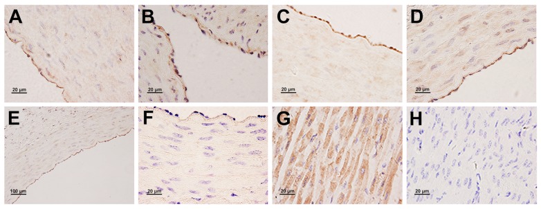
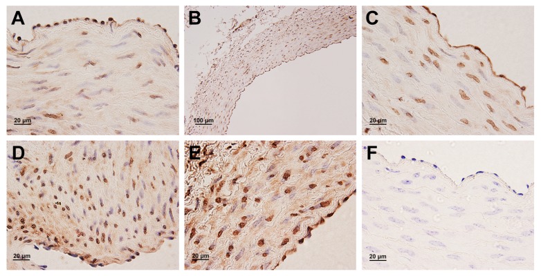
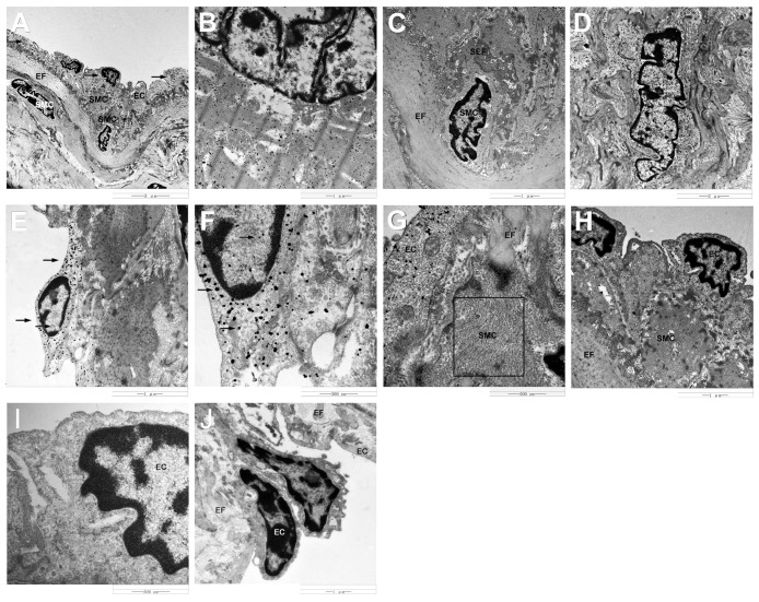
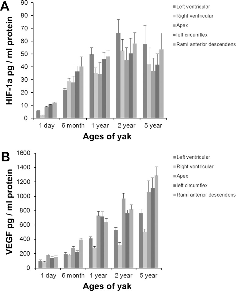
The study aimed to identify the changes of anatomic and microscopic structure and the expression and localization of hypoxia-inducible factor (HIF)-1α and vascular endothelial growth factor (VEGF) in the myocardium and coronary artery of the yak heart adapted to chronic hypoxia with aging. Thirty-two yaks (1 day, 6 months, 1 year, 2 years, and 5 year old) were included, and immunoelectronmicroscopy, immunohistochemistry, and enzyme-linked immunosorbent assay (ELISA) were used. Right ventricular hypertrophy was not present in yaks with aging. There was no intima thickening phenomenon in the coronary artery. The ultrastructure of myofibrils, mitochondria, and collagen fibers and the diameter and quantity of collagen changed significantly with aging. The enzymatic activity of complexes I, II, and V increased with age. Immunogold labeling showed the localization of HIF-1α protein in the cytoplasm and nuclei of endothelial cells and cytoplasm of cardiac muscle cells, and VEGF protein in the nuclei and perinuclei areas of smooth muscle cells of coronary artery, and in the cytoplasm and nuclei of endothelial cells. ELISA results showed that HIF-1α secretion significantly increased in the myocardium and coronary artery from an age of 1 day to 2 years of yaks and decreased in old yaks. However, VEGF protein always increased with aging. The findings of this study suggest that 6 months is a key age of yak before which there are some adaptive changes to deal with low-oxygen environment, and there is a maturation of the yak heart from the age of 6 months to 2 years.
本研究旨在确定随着年龄增长适应慢性缺氧的牦牛心脏心肌和冠状动脉的解剖及微观结构变化,以及缺氧诱导因子(HIF)-1α和血管内皮生长因子(VEGF)的表达与定位。研究纳入了32头牦牛(1日龄、6月龄、1岁、2岁和5岁),并采用了免疫电子显微镜、免疫组织化学和酶联免疫吸附测定(ELISA)方法。随着年龄增长,牦牛未出现右心室肥大。冠状动脉无内膜增厚现象。肌原纤维、线粒体和胶原纤维的超微结构以及胶原的直径和数量随年龄显著变化。复合物I、II和V的酶活性随年龄增加。免疫金标记显示HIF-1α蛋白定位于内皮细胞的细胞质和细胞核以及心肌细胞的细胞质中,VEGF蛋白定位于冠状动脉平滑肌细胞核及核周区域以及内皮细胞的细胞质和细胞核中。ELISA结果显示,从1日龄到2岁龄的牦牛,心肌和冠状动脉中HIF-1α分泌显著增加,而老龄牦牛中则减少。然而,VEGF蛋白始终随年龄增加。本研究结果表明,6月龄是牦牛的关键年龄,在此之前存在一些适应低氧环境的变化,并且牦牛心脏在6月龄到2岁龄之间成熟。