Yang Hong, Guan Liuyuan, Li Shun, Jiang Ying, Xiong Niya, Li Li, Wu Chunhui, Zeng Hongjuan, Liu Yiyao
Department of Biophysics, School of Life Science and Technology of China, Chengdu 610054, Sichuan, P.R. China.
Center for Information in Biomedicine, University of Electronic Science and Technology of China, Chengdu 610054, Sichuan, P.R. China.
Oncotarget. 2016 Mar 29;7(13):16227-47. doi: 10.18632/oncotarget.7583.
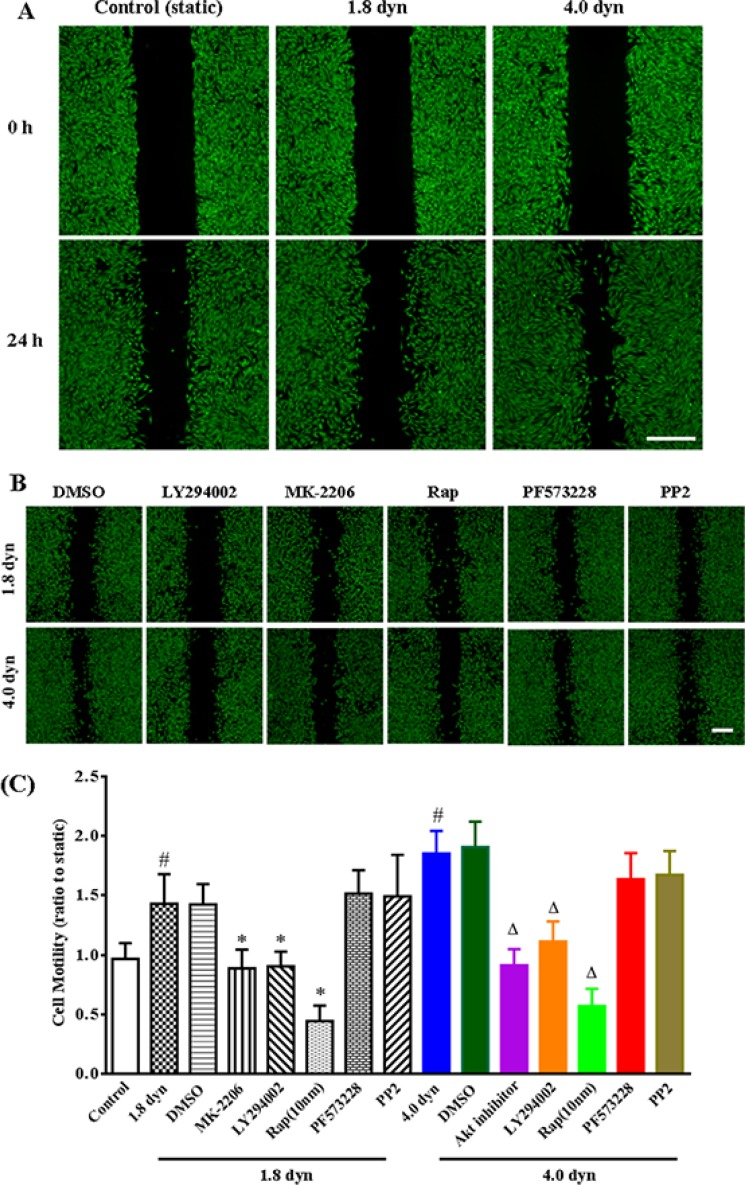
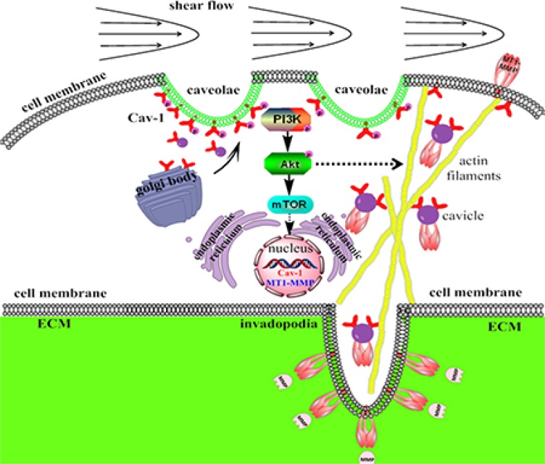
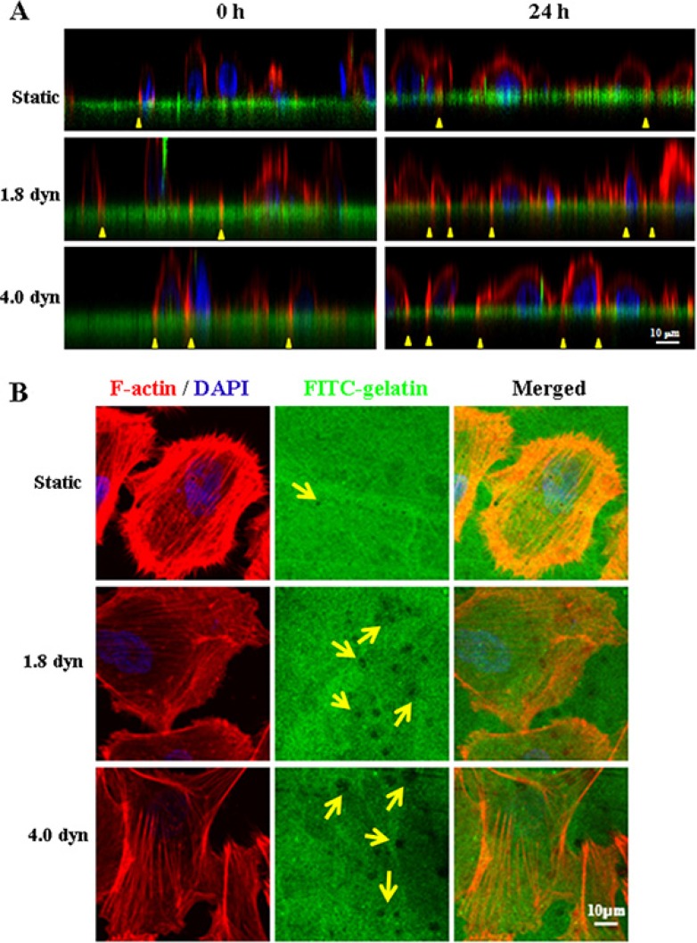
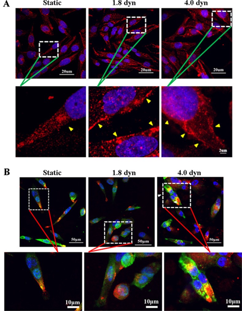
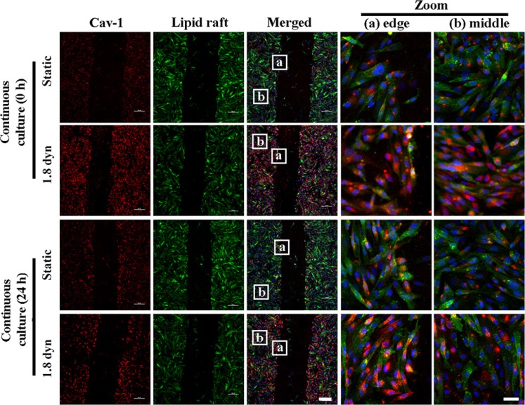
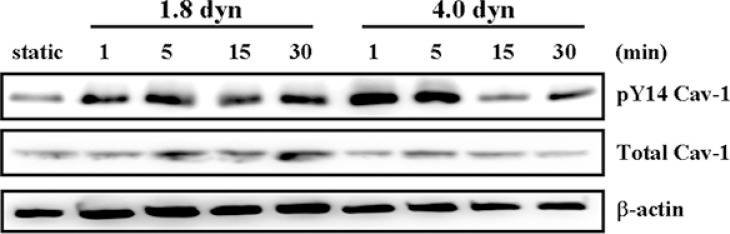
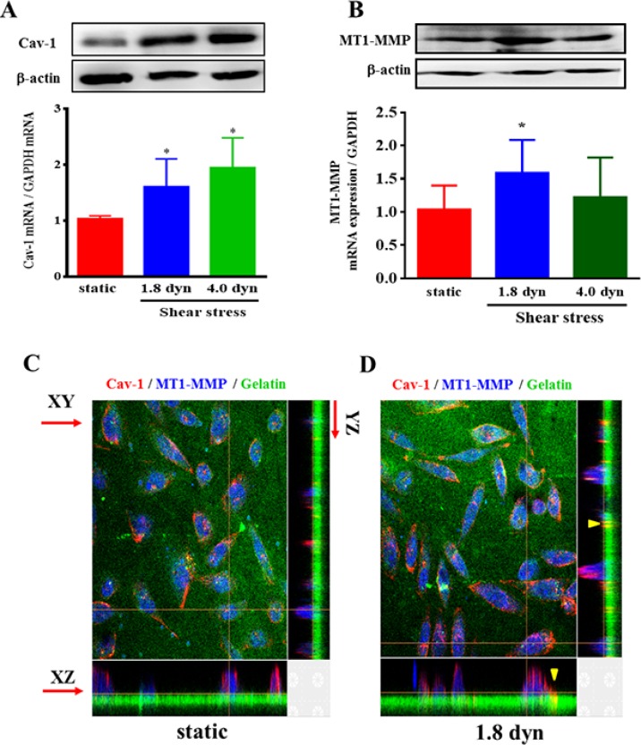
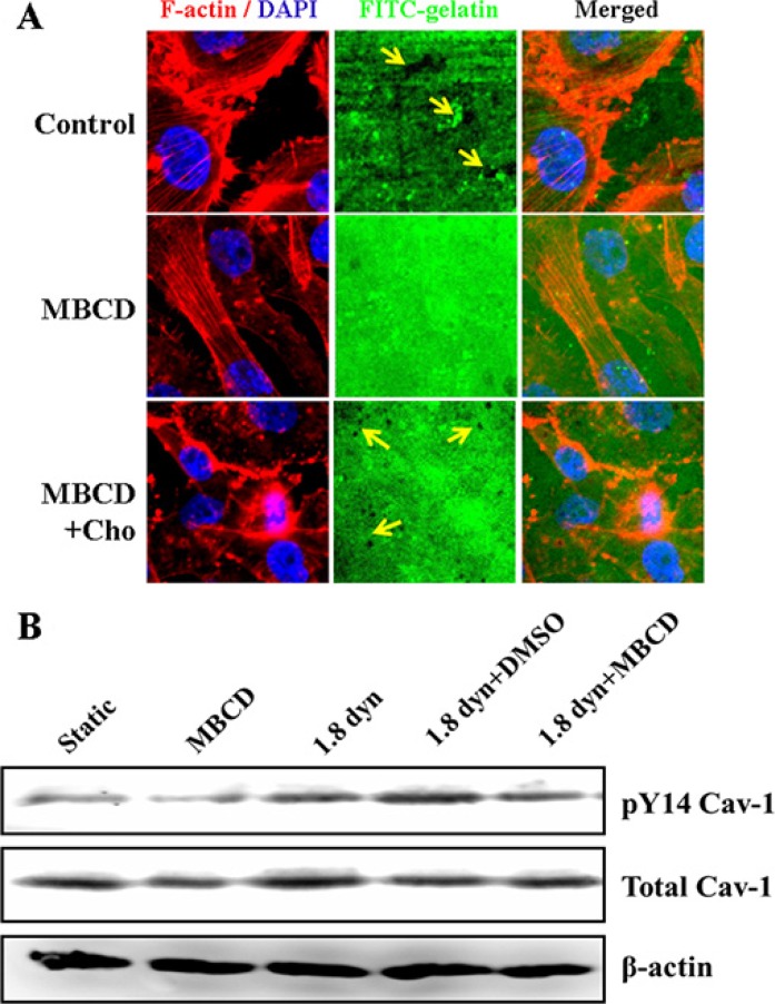
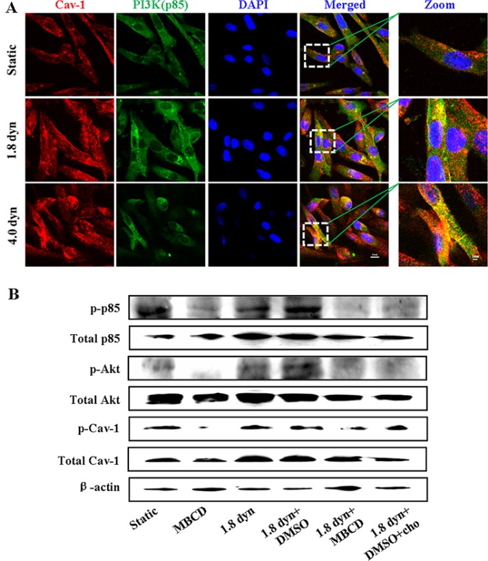
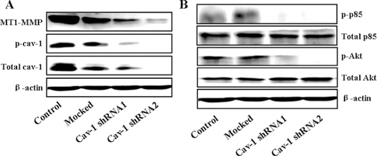
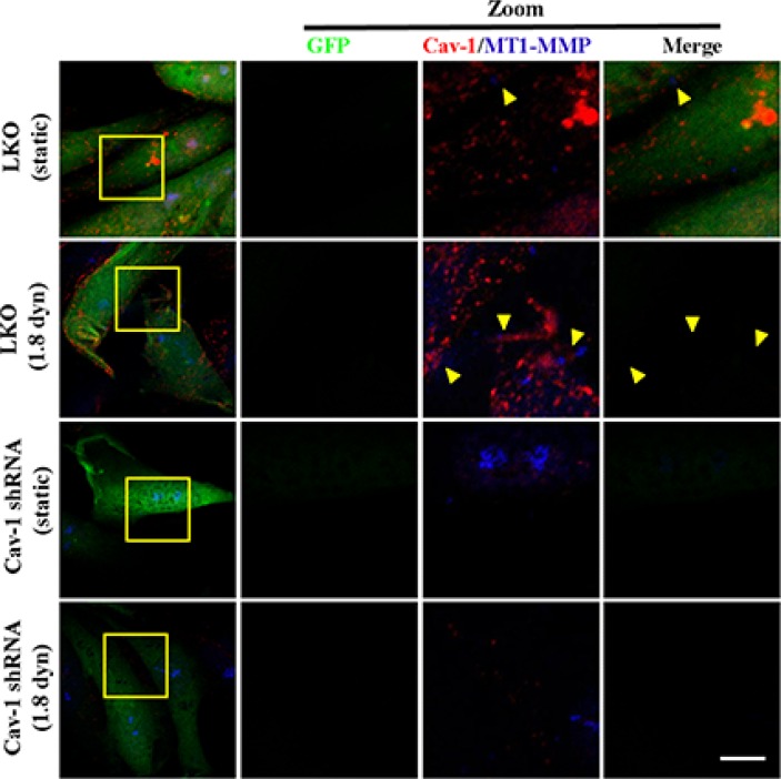
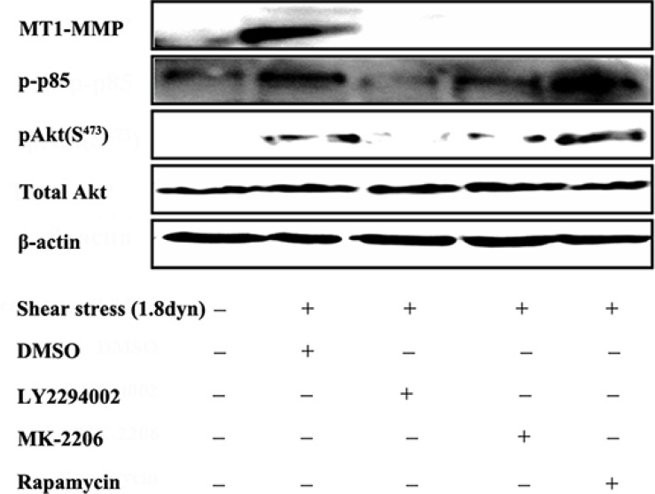
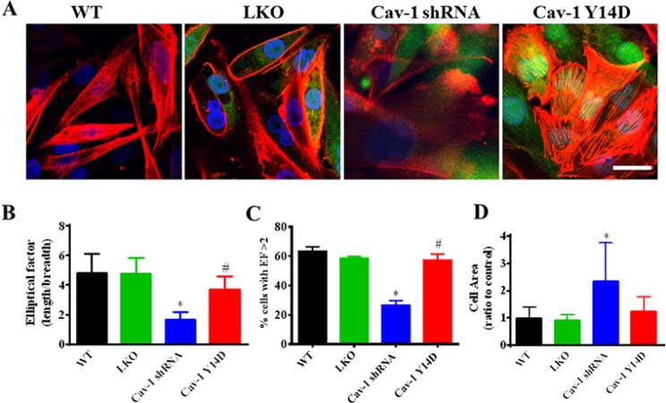
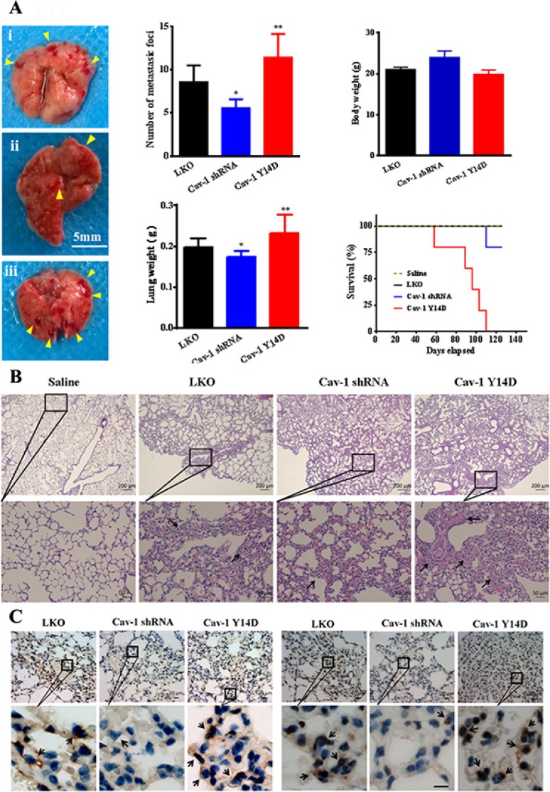
Cancer cells are subjected to fluid shear stress during passage through the venous and lymphatic system. Caveolin-1 (Cav-1), a principal structural component of caveolar membrane domains, contributes to cancer development but its mechanobiological roles under low shear stress (LSS) conditions remain largely unknown. Here, we identified Cav-1 is mechanosensitive to LSS exposure, and its activation-induced PI3K/Akt/mTOR signaling promotes motility, invadopodia formation and metastasis of breast carcinoma MDA-MB-231 cells. Application of LSS (1.8 and 4.0 dynes/cm2) to MDA-MB-231 cells significantly increased the cell motility, invadopodia formation, MT1-MMP expression, ECM degradation, and also induced a sustained activation of Cav-1 and PI3K/Akt/mTOR signaling cascades. Methyl-β-cyclodextrin-caused caveolae destruction markedly decreased LSS-induced activation of both Cav-1 and PI3K/Akt/mTOR, leading to suppress MT1-MMP expression, inhibit invadopodia formation and ECM degradation, suggesting that caveolae integrity also involved in metastasis. Immunocytochemical assay showed that LSS induces the Cav-1 clustering in lipid rafts and co-localization of Cav-1 and MT1-MMP on invadopodia. Immunofluorescence confocal analysis demonstrated that Cav-1 activation were required for the acquisition of a polarized phenotype in MDA-MB-231 cells. Finally, Cav-1 knockdown significantly suppressed tumor colonization in the lungs and distant metastases in animal models. Our findings highlight the importance of Cav-1 in hematogenous metastasis, and provide new insights into the underlying mechanisms of mechanotransduction induced by LSS.
癌细胞在通过静脉和淋巴系统时会受到流体剪切应力的作用。小窝蛋白-1(Cav-1)是小窝膜结构域的主要结构成分,对癌症发展有促进作用,但其在低剪切应力(LSS)条件下的机械生物学作用仍不清楚。在此,我们发现Cav-1对LSS暴露具有机械敏感性,其激活诱导的PI3K/Akt/mTOR信号传导促进了乳腺癌MDA-MB-231细胞的运动性、侵袭伪足形成和转移。对MDA-MB-231细胞施加LSS(1.8和4.0达因/平方厘米)显著增加了细胞运动性、侵袭伪足形成、MT1-MMP表达、细胞外基质降解,还诱导了Cav-1和PI3K/Akt/mTOR信号级联的持续激活。甲基-β-环糊精导致的小窝破坏显著降低了LSS诱导的Cav-1和PI3K/Akt/mTOR的激活,导致MT1-MMP表达受到抑制,侵袭伪足形成和细胞外基质降解受到抑制,这表明小窝完整性也参与转移过程。免疫细胞化学分析表明,LSS诱导Cav-1在脂筏中聚集以及Cav-1与MT1-MMP在侵袭伪足上共定位。免疫荧光共聚焦分析表明,Cav-1激活是MDA-MB-231细胞获得极化表型所必需的。最后,在动物模型中,Cav-1基因敲低显著抑制了肺部肿瘤定植和远处转移。我们的研究结果突出了Cav-1在血行转移中的重要性,并为LSS诱导的机械转导潜在机制提供了新的见解。