Rombouts Yoann, Jónasdóttir Hulda S, Hipgrave Ederveen Agnes L, Reiding Karli R, Jansen Bas C, Freysdottir Jona, Hardardottir Ingibjörg, Ioan-Facsinay Andreea, Giera Martin, Wuhrer Manfred
Center for Proteomics and Metabolomics, Leiden University Medical Center, Leiden, The Netherlands.
Department of Rheumatology, Leiden University Medical Center, Leiden, The Netherlands.
Glycoconj J. 2016 Jun;33(3):457-70. doi: 10.1007/s10719-015-9648-9. Epub 2016 Feb 29.
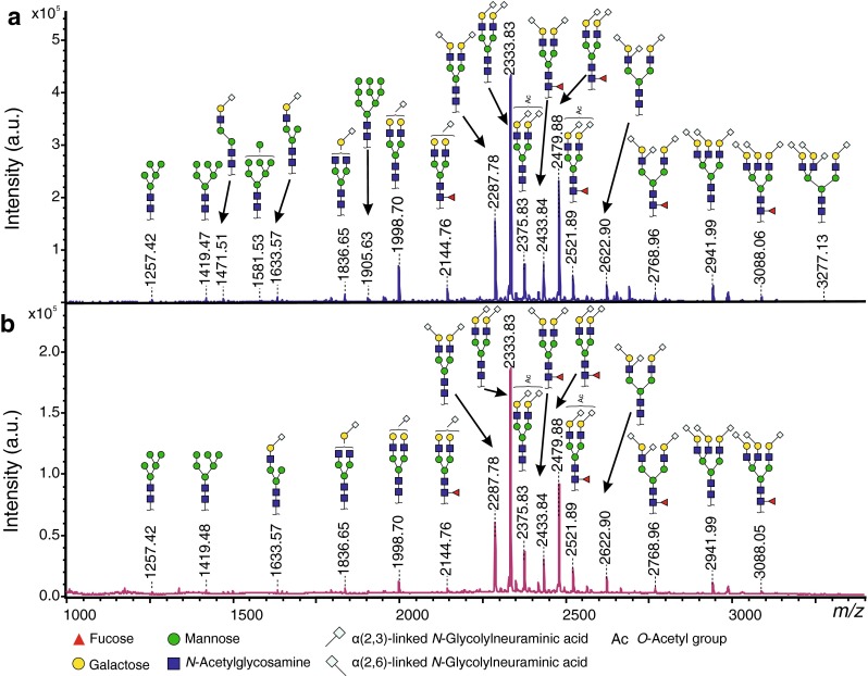
Murine zymosan-induced peritonitis is a widely used model for studying the molecular and cellular events responsible for the initiation, persistence and/or resolution of inflammation. Among these events, it is becoming increasingly evident that changes in glycosylation of proteins, especially in the plasma and at the site of inflammation, play an important role in the inflammatory response. Using matrix-assisted laser desorption/ionization time-of-flight mass spectrometry (MALDI-TOF-MS)-based glycosylation profiling, we investigated the qualitative and quantitative effect of zymosan-induced peritonitis on N-glycosylation in mouse plasma and peritoneal fluid. Our results show that both N-glycomes exhibit highly similar glycosylation patterns, consisting mainly of diantennary and triantennary complex type N-glycans with high levels (>95 %) of galactosylation and sialylation (mostly NeuGc) and a medium degree of core fucosylation (30 %). Moreover, MS/MS structural analysis, assisted by linkage-specific derivatization of sialic acids, revealed the presence of O-acetylated sialic acids as well as disialylated antennae ("branching sialylation") characterized by the presence of α2-6-linked NeuGc on the GlcNAc of the NeuGcα2-3-Galβ1-3-GlcNAc terminal motif. A significant decrease of (core) fucosylation together with an increase of both α2-3-linked NeuGc and "branching sialylation" were observed in N-glycomes of mice challenged with zymosan, but not in control mice injected with PBS. Importantly, substantial changes in glycosylation were already observed 12 h after induction of peritonitis, thereby demonstrating an unexpected velocity of the biological mechanisms involved.
小鼠酵母聚糖诱导的腹膜炎是一种广泛用于研究炎症起始、持续和/或消退相关分子和细胞事件的模型。在这些事件中,越来越明显的是,蛋白质糖基化的变化,尤其是在血浆和炎症部位的变化,在炎症反应中起重要作用。我们使用基于基质辅助激光解吸/电离飞行时间质谱(MALDI-TOF-MS)的糖基化分析方法,研究了酵母聚糖诱导的腹膜炎对小鼠血浆和腹腔液中N-糖基化的定性和定量影响。我们的结果表明,两种N-糖组均呈现高度相似的糖基化模式,主要由双天线和三天线复合型N-聚糖组成,具有高水平(>95%)的半乳糖基化和唾液酸化(主要是NeuGc)以及中等程度的核心岩藻糖基化(30%)。此外,通过唾液酸的连接特异性衍生化辅助的MS/MS结构分析,揭示了O-乙酰化唾液酸以及双唾液酸化天线(“分支唾液酸化”)的存在,其特征是在NeuGcα2-3-Galβ1-3-GlcNAc末端基序的GlcNAc上存在α2-6连接的NeuGc。在用酵母聚糖攻击的小鼠的N-糖组中观察到(核心)岩藻糖基化显著降低,同时α2-3连接的NeuGc和“分支唾液酸化”均增加,但在注射PBS的对照小鼠中未观察到。重要的是,在腹膜炎诱导后12小时就已经观察到糖基化的实质性变化,从而证明了所涉及的生物学机制具有意想不到的速度。