• 文献检索
  • 文档翻译
  • 深度研究
  • 学术资讯
  • Suppr Zotero 插件Zotero 插件
  • 邀请有礼
  • 套餐&价格
  • 历史记录
应用&插件
Suppr Zotero 插件Zotero 插件浏览器插件Mac 客户端Windows 客户端微信小程序
定价
高级版会员购买积分包购买API积分包
服务
文献检索文档翻译深度研究API 文档MCP 服务
关于我们
关于 Suppr公司介绍联系我们用户协议隐私条款
关注我们

Suppr 超能文献

核心技术专利:CN118964589B侵权必究
粤ICP备2023148730 号-1Suppr @ 2026

文献检索

告别复杂PubMed语法,用中文像聊天一样搜索,搜遍4000万医学文献。AI智能推荐,让科研检索更轻松。

立即免费搜索

文件翻译

保留排版,准确专业,支持PDF/Word/PPT等文件格式,支持 12+语言互译。

免费翻译文档

深度研究

AI帮你快速写综述,25分钟生成高质量综述,智能提取关键信息,辅助科研写作。

立即免费体验

利用培养的角膜缘活检外植体的三级生长物自体移植在兔角膜缘干细胞缺乏模型中的角膜恢复情况

Corneal recovery in a rabbit limbal stem cell deficiency model by autologous grafts of tertiary outgrowths from cultivated limbal biopsy explants.

作者信息

Selver Ozlem Barut, Durak Ismet, Gürdal Mehmet, Baysal Kemal, Ates Halil, Ozbek Zeynep, Wang Zheng, Wu Albert, Wolosin J Mario

机构信息

Department of Ophtalmology, Dokuz Eylul University School of Medicine, Izmir, Turkey.

Department of Biochemistry, Dokuz Eylul University School of Medicine, Izmir, Turkey.

出版信息

Mol Vis. 2016 Feb 11;22:138-49. eCollection 2016.

PMID:26937166
原文链接:https://pmc.ncbi.nlm.nih.gov/articles/PMC4757454/
Abstract

PURPOSE

To determine the corneal regenerative capacity of sequentially generated primary, secondary, and tertiary limbal explant outgrowths in a limbal stem cell deficiency (LSCD) surgical model.

METHODS

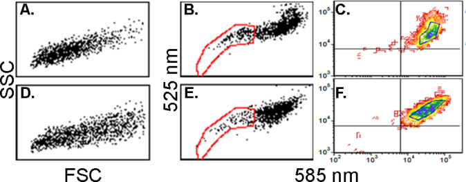
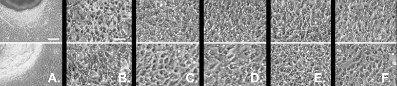
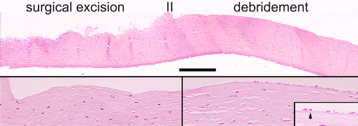
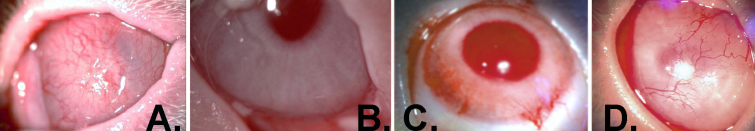
Two-millimeter-long limbal shallow biopsies were surgically excised from the upper quadrant of the right eye of rabbits and set on preserved amniotic membrane for explant culture. After the generation of primary outgrowth, the biopsies were sequentially transferred to new amniotic membrane to generate secondary and then tertiary outgrowths. Eighteen rabbits were subjected to a 360° limbal peritomy extending into the scleral zone and combined with superficial keratectomy of the corneal periphery and thorough mechanical debridement of the central cornea in their left eye. Right eye outgrowths, six of each generation, were engrafted on the ocular surface. Clinical outcomes (neovascularization, corneal clarity, and corneal fluorescein staining) were graded after 6 months. Post-mortem corneas were compared with histology, immunochemistry for p63 and Krt3, ABCG2-dependent dye exclusion, and capacity for outgrowths in explant culture.

RESULTS

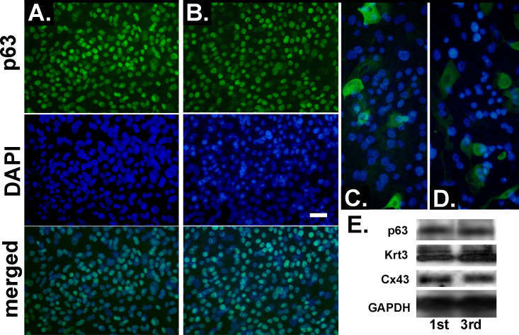
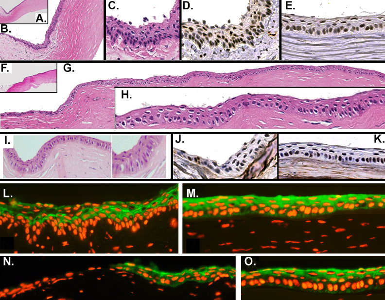
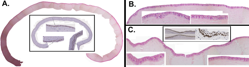
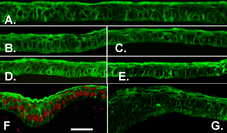
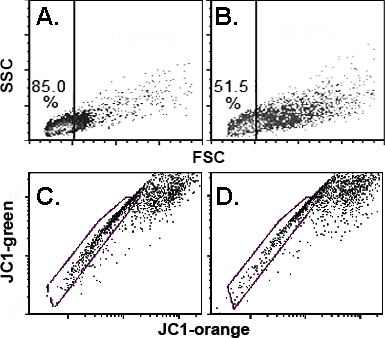
Immunohistology and western blot of the outgrowths for p63 and Krt3 indicated no differences in expression between the primary and tertiary outgrowths for these two markers of growth and differentiation. Clinically, all rabbits treated with amniotic membrane alone developed severe LSCD. Most rabbits grafted with cell outgrowths from all three outgrowth generations achieved stable (>6 months) recovery of the ocular surface. There were partial failures of grafts performed with two secondary and tertiary outgrowths. However, Kruskal-Wallis statistical analysis of the clinical scores yielded no significant difference between the three groups (p=0.524). Histology showed full anatomic recovery of grafts made with primary and tertiary outgrowths. Krt3 and p63 expression throughout the whole limbal corneal epithelium with primary or tertiary outgrowths was not distinguishable from each other. The percentage of dye-excluding cells present within this zone and the capacity of the explant epithelial outgrowth of the regenerated peripheral corneal zone were also on par with those of the donor corneas. The Krt3-negative cells that characterize the basal epithelial layer of the normal limbus could not be found in any regenerated cornea from the primary to tertiary outgrowths.

CONCLUSIONS

Our results demonstrate that in rabbits post-primary explant outgrowths retain the capacity for LSCD recovery found in primary explants.

摘要

目的

在角膜缘干细胞缺乏(LSCD)手术模型中,确定依次生成的原代、二代和三代角膜缘外植体生长物的角膜再生能力。

方法

从兔右眼上象限手术切除2毫米长的角膜缘浅层活检组织,置于保存的羊膜上进行外植体培养。原代生长物生成后,将活检组织依次转移到新的羊膜上,生成二代和三代生长物。18只兔子左眼进行360°角膜缘环行切除术并延伸至巩膜区,同时联合角膜周边浅层角膜切除术及中央角膜彻底机械清创术。将右眼各代的6个生长物移植到眼表。6个月后对临床结果(新生血管形成、角膜透明度和角膜荧光素染色)进行分级。对死后的角膜进行组织学、p63和角蛋白3(Krt3)免疫化学、ABCG2依赖性染料排除以及外植体培养中的生长能力比较。

结果

对生长物进行p63和Krt3的免疫组织化学和蛋白质印迹分析表明,这两种生长和分化标志物在原代和三代生长物中的表达没有差异。临床上,所有仅接受羊膜治疗的兔子均发生严重的LSCD。大多数移植了三代生长物细胞的兔子实现了眼表稳定(>6个月)恢复。二代和三代生长物的移植有部分失败情况。然而,对临床评分进行Kruskal-Wallis统计分析显示,三组之间无显著差异(p=0.524)。组织学显示,原代和三代生长物移植后的解剖结构完全恢复。原代或三代生长物的整个角膜缘上皮中Krt3和p63的表达彼此无明显差异。该区域内染料排除细胞的百分比以及再生周边角膜区外植体上皮生长能力也与供体角膜相当。在从原代到三代生长物的任何再生角膜中均未发现正常角膜缘基底上皮层特有的Krt3阴性细胞。

结论

我们的结果表明,在兔子中,原代外植体生长物之后的生长物保留了原代外植体中发现的LSCD恢复能力。

https://cdn.ncbi.nlm.nih.gov/pmc/blobs/b3c8/4757454/2a21080887bb/mv-v22-138-f9.jpg
https://cdn.ncbi.nlm.nih.gov/pmc/blobs/b3c8/4757454/162c048c4a45/mv-v22-138-f1.jpg
https://cdn.ncbi.nlm.nih.gov/pmc/blobs/b3c8/4757454/f7bc3cec99ac/mv-v22-138-f2.jpg
https://cdn.ncbi.nlm.nih.gov/pmc/blobs/b3c8/4757454/e6c8aca45d24/mv-v22-138-f3.jpg
https://cdn.ncbi.nlm.nih.gov/pmc/blobs/b3c8/4757454/5f4394a07c40/mv-v22-138-f4.jpg
https://cdn.ncbi.nlm.nih.gov/pmc/blobs/b3c8/4757454/4026d31c6f18/mv-v22-138-f5.jpg
https://cdn.ncbi.nlm.nih.gov/pmc/blobs/b3c8/4757454/3aefefae3247/mv-v22-138-f6.jpg
https://cdn.ncbi.nlm.nih.gov/pmc/blobs/b3c8/4757454/95043161577a/mv-v22-138-f7.jpg
https://cdn.ncbi.nlm.nih.gov/pmc/blobs/b3c8/4757454/2eb39b28b051/mv-v22-138-f8.jpg
https://cdn.ncbi.nlm.nih.gov/pmc/blobs/b3c8/4757454/2a21080887bb/mv-v22-138-f9.jpg

相似文献

1
Corneal recovery in a rabbit limbal stem cell deficiency model by autologous grafts of tertiary outgrowths from cultivated limbal biopsy explants.利用培养的角膜缘活检外植体的三级生长物自体移植在兔角膜缘干细胞缺乏模型中的角膜恢复情况
Mol Vis. 2016 Feb 11;22:138-49. eCollection 2016.
2
Factors affecting outcome following transplantation of ex vivo expanded limbal epithelium on amniotic membrane for total limbal deficiency in rabbits.影响兔完全角膜缘缺陷模型中羊膜上体外扩增角膜缘上皮移植术后结局的因素
Invest Ophthalmol Vis Sci. 2002 Aug;43(8):2584-92.
3
Characterization of the corneal surface in limbal stem cell deficiency and after transplantation of cultivated limbal epithelium.角膜缘干细胞缺乏症及培养角膜缘上皮移植后的角膜表面特征
Ophthalmology. 2009 Jun;116(6):1048-56. doi: 10.1016/j.ophtha.2009.01.005. Epub 2009 Apr 25.
4
Functional reconstruction of rabbit corneal epithelium by human limbal cells cultured on amniotic membrane.羊膜上培养的人角膜缘细胞对兔角膜上皮的功能重建
Mol Vis. 2003 Dec 8;9:635-43.
5
Loss of corneal epithelial stem cell properties in outgrowths from human limbal explants cultured on intact amniotic membrane.在完整羊膜上培养的人角膜缘外植体长出物中角膜上皮干细胞特性的丧失。
Regen Med. 2008 May;3(3):329-42. doi: 10.2217/17460751.3.3.329.
6
[Transplantation of bioengineered corneal epithelium for the treatment of total limbal stem cell deficiency in rabbit].生物工程角膜上皮移植治疗兔完全性角膜缘干细胞缺乏症
Zhonghua Yan Ke Za Zhi. 2006 Aug;42(8):679-85.
7
Cell delivery with fixed amniotic membrane reconstructs corneal epithelium in rabbits with limbal stem cell deficiency.固定羊膜细胞移植修复兔角膜缘干细胞缺乏症的角膜上皮。
Invest Ophthalmol Vis Sci. 2011 Feb 9;52(2):724-30. doi: 10.1167/iovs.10-5291.
8
Regeneration of corneal epithelium utilizing a collagen vitrigel membrane in rabbit models for corneal stromal wound and limbal stem cell deficiency.利用胶原玻璃体膜在兔角膜基质伤口和角膜缘干细胞缺乏模型中进行角膜上皮再生。
Acta Ophthalmol. 2015 Feb;93(1):e57-66. doi: 10.1111/aos.12503. Epub 2014 Dec 11.
9
Corneal Recovery Following Rabbit Peripheral Blood Mononuclear Cell-Amniotic Membrane Transplantation with Antivascular Endothelial Growth Factor in Limbal Stem Cell Deficiency Rabbits.兔角膜缘干细胞缺乏症外周血单个核细胞-羊膜移植联合抗血管内皮生长因子治疗后角膜的恢复。
Tissue Eng Part C Methods. 2020 Oct;26(10):541-552. doi: 10.1089/ten.TEC.2020.0209.
10
Limbal stem cell and oral mucosal epithelial transplantation from ex vivo cultivation in LSCD-induced rabbits: histology and immunologic study of the transplant epithelial sheet.体外培养的角膜缘干细胞和口腔黏膜上皮移植于兔角膜缘干细胞缺乏诱导模型:移植上皮片的组织学和免疫学研究
Int Ophthalmol. 2017 Dec;37(6):1289-1298. doi: 10.1007/s10792-016-0402-5. Epub 2016 Dec 2.

引用本文的文献

1
Autologous blood in the management of ocular surface disorders.自体血在眼表疾病治疗中的应用
World J Exp Med. 2024 Dec 20;14(4):96412. doi: 10.5493/wjem.v14.i4.96412.
2
Titanium Powder 3D-Printing Technology for a Novel Keratoprosthesis in Alkali-Burned Rabbits.钛粉 3D 打印技术在碱性烧伤兔新型角膜假体中的应用。
Transl Vis Sci Technol. 2022 Aug 1;11(8):14. doi: 10.1167/tvst.11.8.14.
3
Cultivated Autologous Limbal Epithelial Cell Transplantation: New Frontier in the Treatment of Limbal Stem Cell Deficiency.培养自体角膜缘上皮细胞移植:治疗角膜缘干细胞缺陷的新前沿。

本文引用的文献

1
Inhibition of TGFβ cell signaling for limbal explant culture in serumless, defined xeno-free conditions.在无血清、明确的无动物源条件下抑制转化生长因子β(TGFβ)细胞信号通路用于角膜缘外植体培养。
Exp Eye Res. 2016 Apr;145:48-57. doi: 10.1016/j.exer.2015.10.021. Epub 2015 Nov 7.
2
Connecting Mitochondria, Metabolism, and Stem Cell Fate.连接线粒体、新陈代谢与干细胞命运
Stem Cells Dev. 2015 Sep 1;24(17):1957-71. doi: 10.1089/scd.2015.0117. Epub 2015 Jul 2.
3
Cord blood-derived CD34+ hematopoietic cells with low mitochondrial mass are enriched in hematopoietic repopulating stem cell function.
Am J Ophthalmol. 2022 Jul;239:244-268. doi: 10.1016/j.ajo.2022.03.015. Epub 2022 Mar 18.
4
Differentiation of rat adipose-derived mesenchymal stem cells into corneal-like epithelial cells driven by PAX6.由PAX6驱动的大鼠脂肪间充质干细胞向角膜样上皮细胞的分化
Exp Ther Med. 2018 Feb;15(2):1424-1432. doi: 10.3892/etm.2017.5576. Epub 2017 Nov 27.
5
Limbal Stem Cell Deficiency and Treatment with Stem Cell Transplantation.角膜缘干细胞缺乏症与干细胞移植治疗
Turk J Ophthalmol. 2017 Oct;47(5):285-291. doi: 10.4274/tjo.72593. Epub 2017 Oct 27.
6
Application of JC1 for non-toxic isolation of cells with MDR transporter activity by flow cytometry.通过流式细胞术应用JC1对具有多药耐药转运蛋白活性的细胞进行无毒分离。
PLoS One. 2017 Apr 5;12(4):e0174905. doi: 10.1371/journal.pone.0174905. eCollection 2017.
低线粒体质量的脐带血来源 CD34+ 造血细胞富含造血重建造血干细胞功能。
Haematologica. 2013 Jul;98(7):1022-9. doi: 10.3324/haematol.2012.079244. Epub 2013 Jan 24.
4
Ocular surface stem cell transplantation rejection: incidence, characteristics, and outcomes.眼表干细胞移植排斥反应:发生率、特征和结果。
Cornea. 2013 Mar;32(3):229-36. doi: 10.1097/ICO.0b013e318255eac4.
5
Clinical outcomes of xeno-free autologous cultivated limbal epithelial transplantation: a 10-year study.无饲养层自体培养角膜缘上皮移植的临床疗效:一项 10 年研究。
Br J Ophthalmol. 2011 Nov;95(11):1525-9. doi: 10.1136/bjophthalmol-2011-300352. Epub 2011 Sep 2.
6
ABCG2-dependent dye exclusion activity and clonal potential in epithelial cells continuously growing for 1 month from limbal explants.从角膜缘外植体连续培养 1 个月的上皮细胞中 ABCG2 依赖性染料排除活性和克隆潜能。
Invest Ophthalmol Vis Sci. 2011 Jun 17;52(7):4330-7. doi: 10.1167/iovs.10-5897.
7
p63 in epithelial survival, germ cell surveillance, and neoplasia.p63 在上皮细胞存活、生殖细胞监测和肿瘤发生中起作用。
Annu Rev Pathol. 2010;5:349-71. doi: 10.1146/annurev-pathol-121808-102117.
8
One-year follow-up of epithelial corneal cell sheet allografts mounted on platelet poor plasma in rabbits.兔角膜上皮细胞片移植于少血小板血浆的一年随访研究
Mol Vis. 2009 Dec 15;15:2771-9.
9
Autologous transplantation of conjunctival epithelial cells cultured on amniotic membrane in a rabbit model.兔模型中羊膜培养的结膜上皮细胞自体移植
Mol Vis. 2007 Jul 13;13:1138-43.
10
Human amniotic epithelial cells as novel feeder layers for promoting ex vivo expansion of limbal epithelial progenitor cells.人羊膜上皮细胞作为促进角膜缘上皮祖细胞体外扩增的新型饲养层。
Stem Cells. 2007 Aug;25(8):1995-2005. doi: 10.1634/stemcells.2006-0677. Epub 2007 May 10.