Sun Jing, Liu Wei-Hua, Deng Feng-Mei, Luo Yong-Hui, Wen Ke, Zhang Hong, Liu Hai-Rong, Wu Jiang, Su Bing-Yin, Liu Yi-Lun
Department of Neurobiology, Chongqing Key Laboratory of Neurobiology, Third Military Medical University, Chongqing 400038, P.R. China.
Department of Regeneration Key Lab of Sichuan Province, Chengdu Medical College, Chengdu, Sichuan 610500, P.R. China.
Exp Ther Med. 2018 Feb;15(2):1424-1432. doi: 10.3892/etm.2017.5576. Epub 2017 Nov 27.
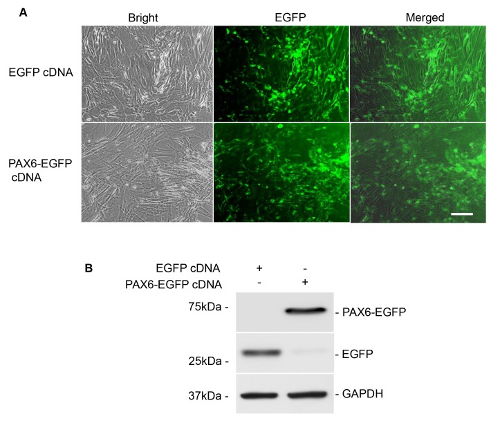
Corneal integrity, transparency and vision acuity are maintained by corneal epithelial cells (CECs), which are continuously renewed by corneal limbal stem cells (LSCs). Deficiency of CECs and/or LSCs is associated with numerous ocular diseases. Paired box (PAX)6 is an eye development-associated transcription factor that is necessary for cell fate determination and differentiation of LSCs and CECs. In the present study, the PAX6 gene was introduced into adipose-derived rat mesenchymal stem cells (ADMSCs) to investigate whether PAX6-transfected cells were able to transdifferentiate into corneal-like epithelial cells and to further verify whether the cells were suitable as a cell source for corneal transplantation. The ADMSCs were isolated from the bilateral inguinal region of healthy Sprague Dawley rats. The characteristics of ADMSCs were identified using flow cytometric analysis. After subculture, ADMSCs underwent transfection with recombinant plasmid containing either PAX6-enhanced green fluorescent protein (EGFP) complementary (c)DNA or EGFP cDNA (blank plasmid group), followed by selection with G418 and determination of the transfection efficiency. Subsequently, the morphology of the ADMSCs and the expression profiles of corneal-specific markers CK3/12 and epithelial-specific adhesion protein were determined. E-cadherin was detected using immunofluorescence staining and western blot analysis at 21 days following transfection. An MTT cell proliferation and a colony formation assay were performed to assess the proliferative activity and clonogenicity of PAX6-transfected ADMSCs. Finally, the PAX6-expressing ADMSCs were transplanted onto the cornea of a rabbits with limbal stem cell deficiency (LSCD). At 21 days after transfection, the ADMSCs with PAX6 transfection exhibited a characteristic flagstone-like appearance with assembled corneal-like epithelial cells, and concomitant prominent expression of the corneal-specific markers cytokeratin 3/12 and E-cadherin. Furthermore, the proliferation and colony formation ability of PAX6-overexpressing ADMSCs was significantly retarded. The transplantation experiment indicated that PAX6-reprogramed ADMSCs attached to and replenished the damaged cornea via formation of stratified corneal epithelium. Taken together, these results suggested that conversion of ADMSCs into corneal-like epithelium may be driven by PAX6 transfection, which makes ADMSCs a promising cell candidate for the treatment of LSCD.
角膜上皮细胞(CECs)维持着角膜的完整性、透明度和视力,而角膜缘干细胞(LSCs)不断更新角膜上皮细胞。CECs和/或LSCs的缺乏与多种眼部疾病相关。配对盒(PAX)6是一种与眼睛发育相关的转录因子,对LSCs和CECs的细胞命运决定和分化至关重要。在本研究中,将PAX6基因导入大鼠脂肪间充质干细胞(ADMSCs),以研究PAX6转染的细胞是否能够转分化为角膜样上皮细胞,并进一步验证这些细胞是否适合作为角膜移植的细胞来源。从健康的Sprague Dawley大鼠双侧腹股沟区域分离ADMSCs。使用流式细胞术分析鉴定ADMSCs的特征。传代培养后,ADMSCs用含有PAX6增强绿色荧光蛋白(EGFP)互补(c)DNA或EGFP cDNA(空白质粒组)的重组质粒进行转染,随后用G418筛选并测定转染效率。随后,测定ADMSCs的形态以及角膜特异性标志物CK3/12和上皮特异性粘附蛋白的表达谱。转染后21天,使用免疫荧光染色和蛋白质印迹分析检测E-钙粘蛋白。进行MTT细胞增殖和集落形成试验,以评估PAX6转染的ADMSCs的增殖活性和克隆形成能力。最后,将表达PAX6的ADMSCs移植到患有角膜缘干细胞缺乏症(LSCD)的兔角膜上。转染后21天,转染PAX6的ADMSCs呈现出特征性的铺路石样外观,伴有角膜样上皮细胞聚集,同时角膜特异性标志物细胞角蛋白3/12和E-钙粘蛋白显著表达。此外,过表达PAX6的ADMSCs的增殖和集落形成能力明显受到抑制。移植实验表明,PAX6重编程的ADMSCs通过形成分层角膜上皮附着并补充受损角膜。综上所述,这些结果表明PAX6转染可能驱动ADMSCs向角膜样上皮细胞转化,这使得ADMSCs成为治疗LSCD有前景的细胞候选者。