Siegmund Sören V, Schlosser Monika, Schildberg Frank A, Seki Ekihiro, De Minicis Samuele, Uchinami Hiroshi, Kuntzen Christian, Knolle Percy A, Strassburg Christian P, Schwabe Robert F
Department of Medicine, Columbia University, College of Physicians and Surgeons, New York, New York, United States of America.
Dept. of Medicine I, University of Bonn, Bonn, Germany.
PLoS One. 2016 Mar 3;11(3):e0150893. doi: 10.1371/journal.pone.0150893. eCollection 2016.
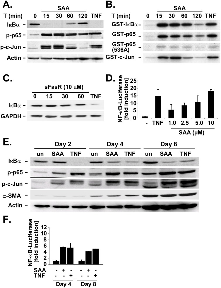
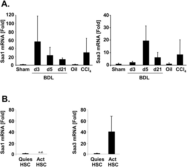
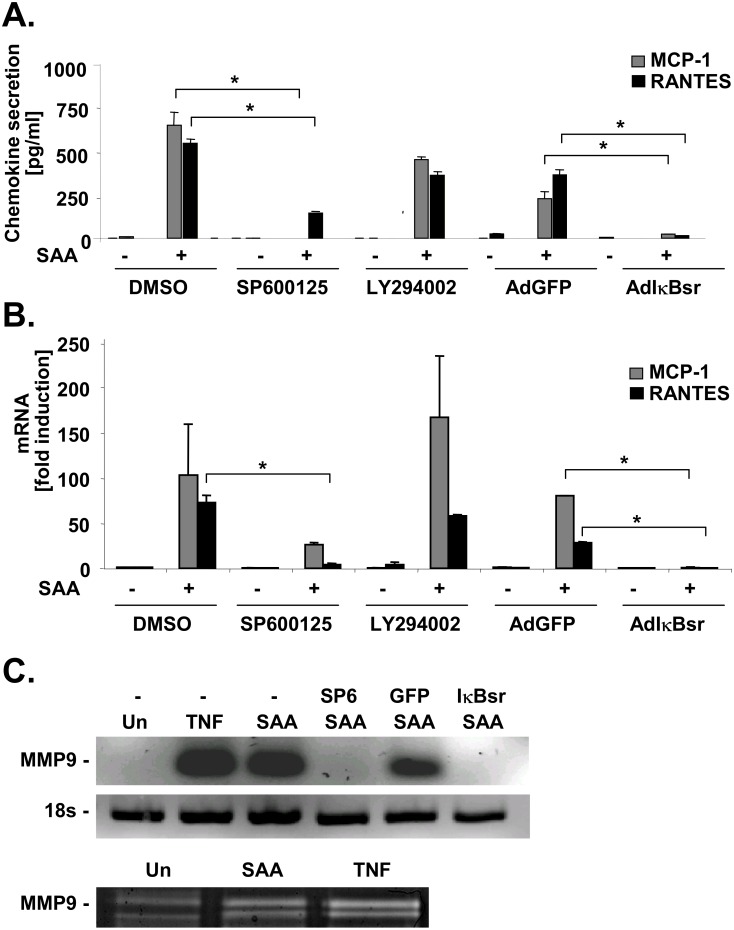
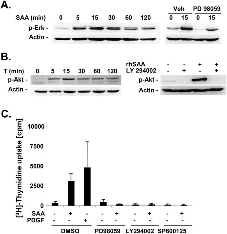
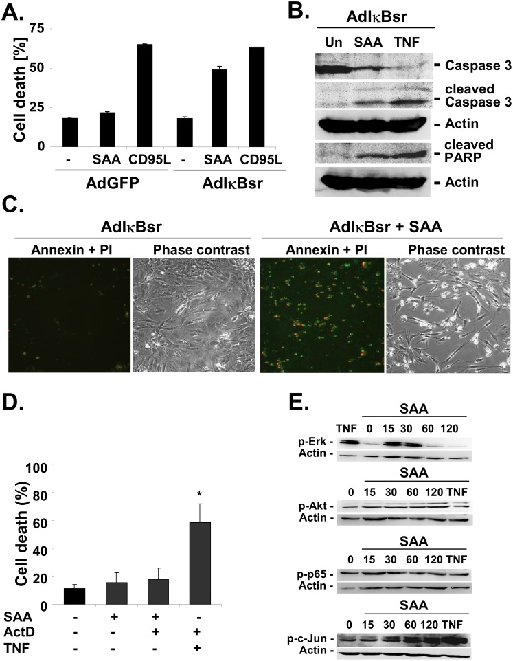
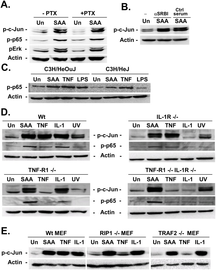
Serum amyloid A (SAA) is an evolutionary highly conserved acute phase protein that is predominantly secreted by hepatocytes. However, its role in liver injury and fibrogenesis has not been elucidated so far. In this study, we determined the effects of SAA on hepatic stellate cells (HSCs), the main fibrogenic cell type of the liver. Serum amyloid A potently activated IκB kinase, c-Jun N-terminal kinase (JNK), Erk and Akt and enhanced NF-κB-dependent luciferase activity in primary human and rat HSCs. Serum amyloid A induced the transcription of MCP-1, RANTES and MMP9 in an NF-κB- and JNK-dependent manner. Blockade of NF-κB revealed cytotoxic effects of SAA in primary HSCs with signs of apoptosis such as caspase 3 and PARP cleavage and Annexin V staining. Serum amyloid A induced HSC proliferation, which depended on JNK, Erk and Akt activity. In primary hepatocytes, SAA also activated MAP kinases, but did not induce relevant cell death after NF-κB inhibition. In two models of hepatic fibrogenesis, CCl4 treatment and bile duct ligation, hepatic mRNA levels of SAA1 and SAA3 were strongly increased. In conclusion, SAA may modulate fibrogenic responses in the liver in a positive and negative fashion by inducing inflammation, proliferation and cell death in HSCs.
血清淀粉样蛋白A(SAA)是一种进化上高度保守的急性期蛋白,主要由肝细胞分泌。然而,其在肝损伤和纤维化形成中的作用迄今尚未阐明。在本研究中,我们确定了SAA对肝星状细胞(HSC)的影响,HSC是肝脏主要的纤维化细胞类型。血清淀粉样蛋白A在原代人源和大鼠HSC中强烈激活IκB激酶、c-Jun氨基末端激酶(JNK)、细胞外信号调节激酶(Erk)和蛋白激酶B(Akt),并增强核因子κB(NF-κB)依赖性荧光素酶活性。血清淀粉样蛋白A以NF-κB和JNK依赖性方式诱导单核细胞趋化蛋白-1(MCP-1)、调节激活正常T细胞表达和分泌因子(RANTES)和基质金属蛋白酶9(MMP9)的转录。NF-κB的阻断揭示了SAA在原代HSC中的细胞毒性作用,伴有细胞凋亡迹象,如半胱天冬酶3和多聚(ADP-核糖)聚合酶(PARP)裂解以及膜联蛋白V染色。血清淀粉样蛋白A诱导HSC增殖,这依赖于JNK、Erk和Akt活性。在原代肝细胞中,SAA也激活丝裂原活化蛋白激酶(MAP激酶),但在NF-κB抑制后未诱导相关细胞死亡。在两种肝纤维化模型,即四氯化碳处理和胆管结扎模型中,SAA1和SAA3的肝脏mRNA水平显著升高。总之,SAA可能通过诱导HSC中的炎症、增殖和细胞死亡,以正负两种方式调节肝脏中的纤维化反应。