Strazielle Nathalie, Creidy Rita, Malcus Christophe, Boucraut José, Ghersi-Egea Jean-François
Brain-i, Lyon, France.
Blood-brain interfaces exploratory platform BIP and FLUID Team, Lyon Neurosciences Research Center, INSERM U1028 CRNS UMR 5292, Université Claude Bernard Lyon-1, Lyon, France.
PLoS One. 2016 Mar 4;11(3):e0150945. doi: 10.1371/journal.pone.0150945. eCollection 2016.
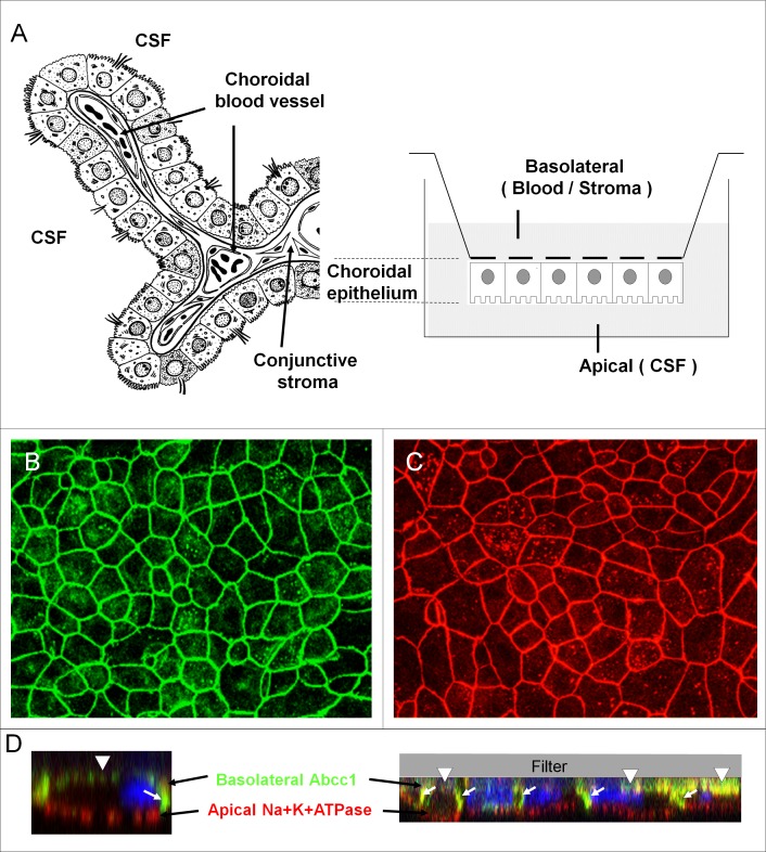
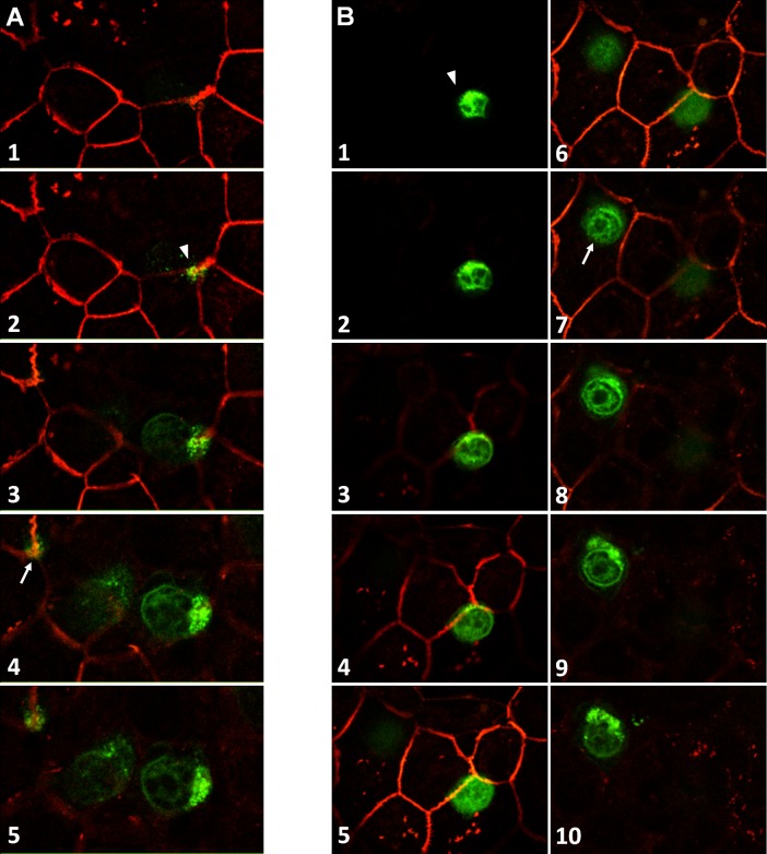
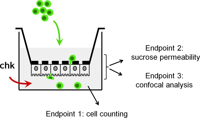
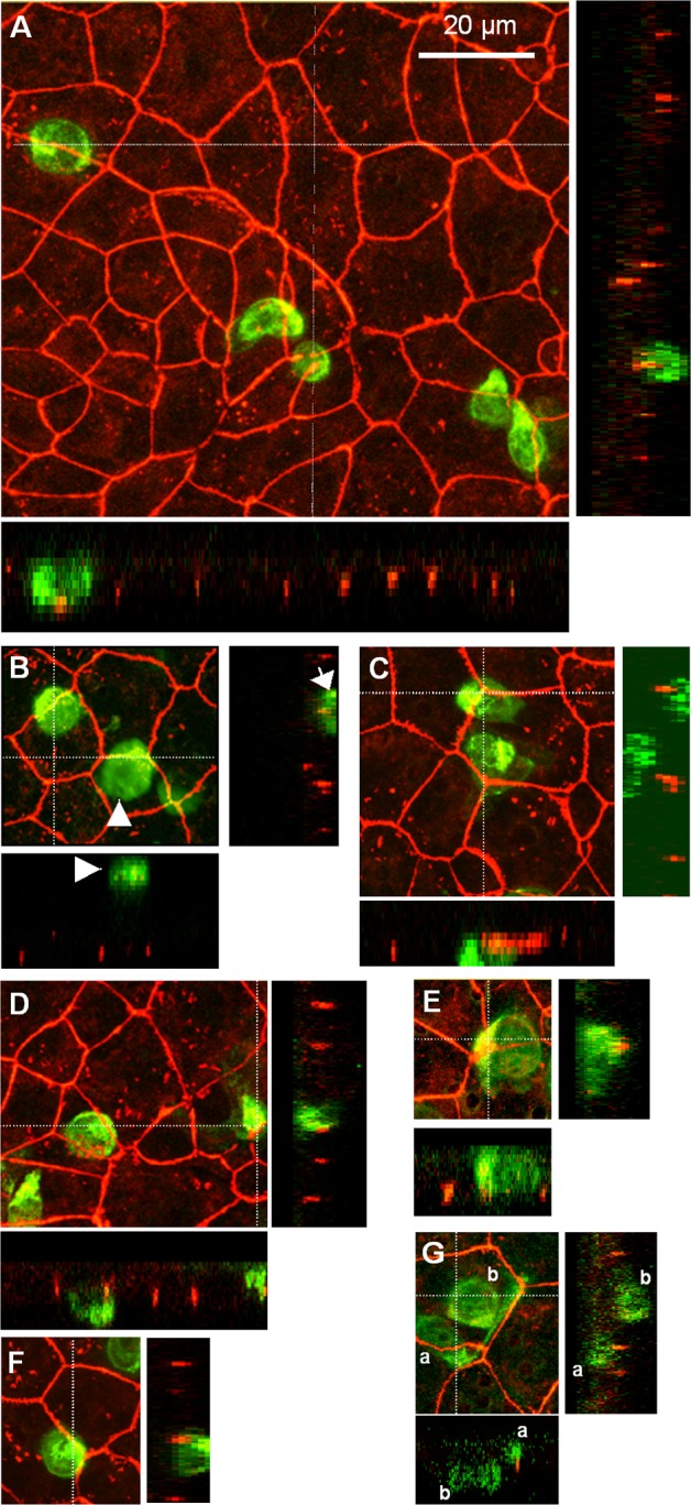
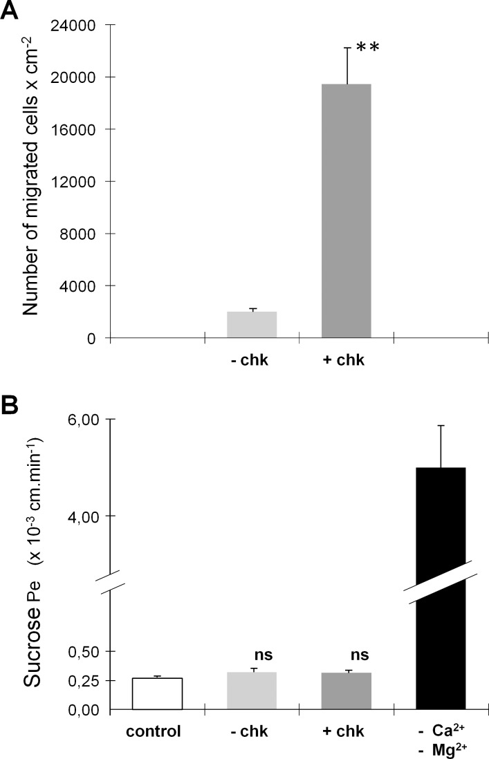
An emerging concept of normal brain immune surveillance proposes that recently and moderately activated central memory T lymphocytes enter the central nervous system (CNS) directly into the cerebrospinal fluid (CSF) via the choroid plexus. Within the CSF space, T cells inspect the CNS environment for cognate antigens. This gate of entry into the CNS could also prevail at the initial stage of neuroinflammatory processes. To actually demonstrate T cell migration across the choroidal epithelium forming the blood-CSF barrier, an in vitro model of the rat blood-CSF barrier was established in an "inverse" configuration that enables cell transmigration studies in the basolateral to apical, i.e. blood/stroma to CSF direction. Structural barrier features were evaluated by immunocytochemical analysis of tight junction proteins, functional barrier properties were assessed by measuring the monolayer permeability to sucrose and the active efflux transport of organic anions. The migratory behaviour of activated T cells across the choroidal epithelium was analysed in the presence and absence of chemokines. The migration pathway was examined by confocal microscopy. The inverse rat BCSFB model reproduces the continuous distribution of tight junction proteins at cell margins, the restricted paracellular permeability, and polarized active transport mechanisms, which all contribute to the barrier phenotype in vivo. Using this model, we present experimental evidence of T cell migration across the choroidal epithelium. Cell migration appears to occur via a paracellular route without disrupting the restrictive barrier properties of the epithelial interface. Apical chemokine addition strongly stimulates T cell migration across the choroidal epithelium. The present data provide evidence for the controlled migration of T cells across the blood-CSF barrier into brain. They further indicate that this recruitment route is sensitive to CSF-borne chemokines, extending the relevance of this migration pathway to neuroinflammatory and neuroinfectious disorders which are typified by elevated chemokine levels in CSF.
正常脑免疫监视的一个新兴概念提出,近期中度激活的中枢记忆T淋巴细胞通过脉络丛直接进入中枢神经系统(CNS)的脑脊液(CSF)。在脑脊液空间内,T细胞检测中枢神经系统环境中的同源抗原。这个进入中枢神经系统的门户在神经炎症过程的初始阶段也可能起主导作用。为了实际证明T细胞穿过形成血脑屏障的脉络膜上皮的迁移,建立了大鼠血脑屏障的体外模型,其呈“反向”构型,能够进行从基底外侧到顶端,即从血液/基质到脑脊液方向的细胞迁移研究。通过紧密连接蛋白的免疫细胞化学分析评估结构屏障特征,通过测量单层对蔗糖的通透性和有机阴离子的主动外排转运评估功能屏障特性。在有和没有趋化因子的情况下分析活化T细胞穿过脉络膜上皮的迁移行为。通过共聚焦显微镜检查迁移途径。反向大鼠血脑屏障模型再现了紧密连接蛋白在细胞边缘的连续分布、限制的细胞旁通透性和极化的主动转运机制,这些都有助于体内的屏障表型。使用这个模型,我们提供了T细胞穿过脉络膜上皮迁移的实验证据。细胞迁移似乎通过细胞旁途径发生,而不会破坏上皮界面的限制性屏障特性。顶端添加趋化因子强烈刺激T细胞穿过脉络膜上皮的迁移。目前的数据为T细胞穿过血脑屏障进入脑内的受控迁移提供了证据。它们进一步表明,这种募集途径对脑脊液中的趋化因子敏感,将这种迁移途径的相关性扩展到以脑脊液中趋化因子水平升高为特征的神经炎症和神经感染性疾病。