Oshida Keiyu, Waxman David J, Corton J Christopher
Integrated Systems Toxicology Division, National Health and Environmental Effects Research Laboratory/ Office of Research and Development, United States Environmental Protection Agency, Research Triangle Park, North Carolina, United States of America.
Division of Cell and Molecular Biology, Department of Biology and Bioinformatics Program, Boston University, Boston, MA 02215, United States of America.
PLoS One. 2016 Mar 9;11(3):e0150284. doi: 10.1371/journal.pone.0150284. eCollection 2016.
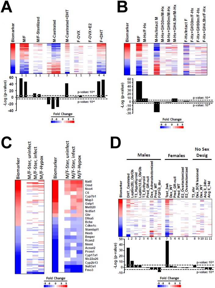
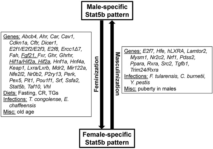
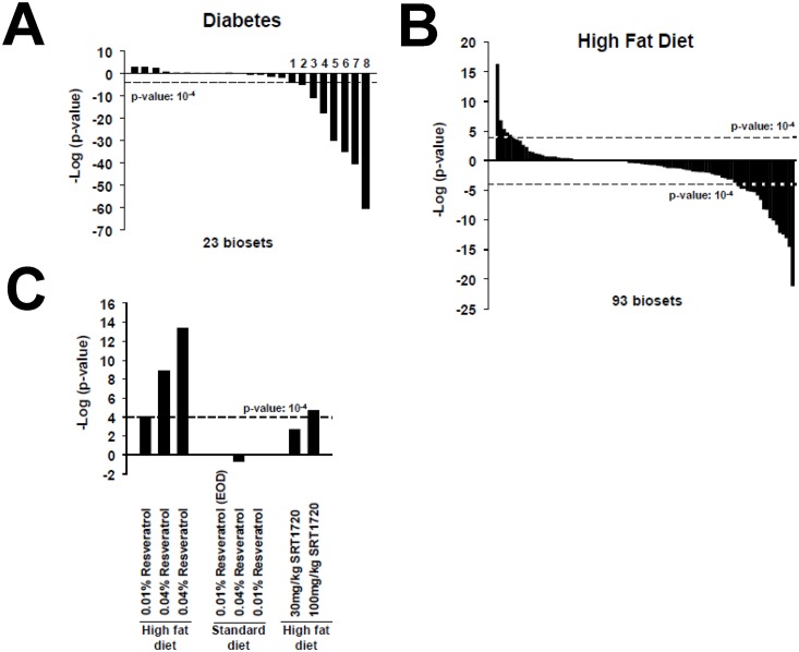
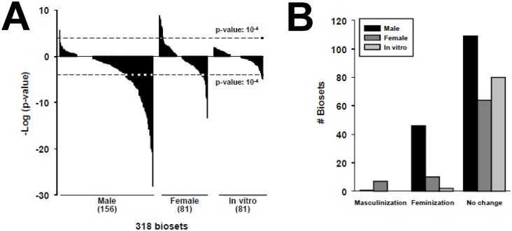
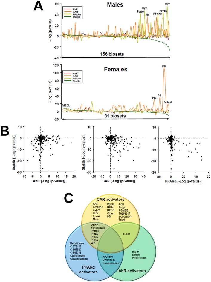
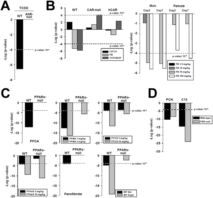
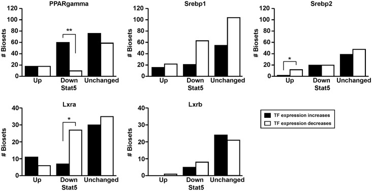
The growth hormone (GH)-activated transcription factor signal transducer and activator of transcription 5b (STAT5b) is a key regulator of sexually dimorphic gene expression in the liver. Suppression of hepatic STAT5b signaling is associated with lipid metabolic dysfunction leading to steatosis and liver cancer. In the companion publication, a STAT5b biomarker gene set was identified and used in a rank-based test to predict both increases and decreases in liver STAT5b activation status/function with high (≥ 97%) accuracy. Here, this computational approach was used to identify chemicals and hormones that activate (masculinize) or suppress (feminize) STAT5b function in a large, annotated mouse liver and primary hepatocyte gene expression compendium. Exposure to dihydrotestosterone and thyroid hormone caused liver masculinization, whereas glucocorticoids, fibroblast growth factor 15, and angiotensin II caused liver feminization. In mouse models of diabetes and obesity, liver feminization was consistently observed and was at least partially reversed by leptin or resveratrol exposure. Chemical-induced feminization of male mouse liver gene expression profiles was a relatively frequent phenomenon: of 156 gene expression biosets from chemically-treated male mice, 29% showed feminization of liver STAT5b function, while <1% showed masculinization. Most (93%) of the biosets that exhibited feminization of male liver were also associated with activation of one or more xenobiotic-responsive receptors, most commonly constitutive activated receptor (CAR) or peroxisome proliferator-activated receptor alpha (PPARα). Feminization was consistently associated with increased expression of peroxisome proliferator-activated receptor gamma (Pparg) but not other lipogenic transcription factors linked to steatosis. GH-activated STAT5b signaling in mouse liver is thus commonly altered by diverse chemicals, and provides a linkage between chemical exposure and dysregulated gene expression associated with adverse effects on the liver.
生长激素(GH)激活的转录因子信号转导子和转录激活子5b(STAT5b)是肝脏中性别二态性基因表达的关键调节因子。肝脏中STAT5b信号传导的抑制与脂质代谢功能障碍有关,可导致脂肪变性和肝癌。在配套论文中,鉴定出了一个STAT5b生物标志物基因集,并将其用于基于排序的测试中,以预测肝脏STAT5b激活状态/功能的增加和降低,准确率高达(≥97%)。在此,这种计算方法被用于在一个大型的、有注释的小鼠肝脏和原代肝细胞基因表达汇编中识别激活(使男性化)或抑制(使女性化)STAT5b功能的化学物质和激素。接触二氢睾酮和甲状腺激素会导致肝脏男性化,而糖皮质激素、成纤维细胞生长因子15和血管紧张素II会导致肝脏女性化。在糖尿病和肥胖症的小鼠模型中,持续观察到肝脏女性化,而瘦素或白藜芦醇暴露至少可部分逆转这种现象。化学诱导的雄性小鼠肝脏基因表达谱女性化是一种相对常见的现象:在来自化学处理雄性小鼠的156个基因表达生物集中,29%显示肝脏STAT5b功能女性化,而<1%显示男性化。大多数(93%)表现出雄性肝脏女性化的生物集也与一种或多种外源性物质反应受体的激活有关,最常见的是组成型激活受体(CAR)或过氧化物酶体增殖物激活受体α(PPARα)。女性化一直与过氧化物酶体增殖物激活受体γ(Pparg)的表达增加有关,但与其他与脂肪变性相关的生脂转录因子无关。因此,小鼠肝脏中GH激活的STAT5b信号传导通常会被多种化学物质改变,并在化学物质暴露与对肝脏产生不利影响的基因表达失调之间建立了联系。