Fernandez-Moure Joseph S, Van Eps Jeffrey L, Rhudy Jessica R, Cabrera Fernando J, Acharya Ghanashyam S, Tasciotti Ennio, Sakamoto Jason, Nichols Joan E
Department of Surgery, Houston Methodist Hospital, Houston, TX, USA; Surgical Advanced Technologies Lab, Department of Biomimetic and Regenerative Medicine, Houston Methodist Research Institute, Houston, TX, USA.
Department of Nanomedicine, Houston Methodist Research Institute, Houston, TX, USA.
J Tissue Eng. 2016 Feb 1;7:2041731415626018. doi: 10.1177/2041731415626018. eCollection 2016 Jan-Dec.
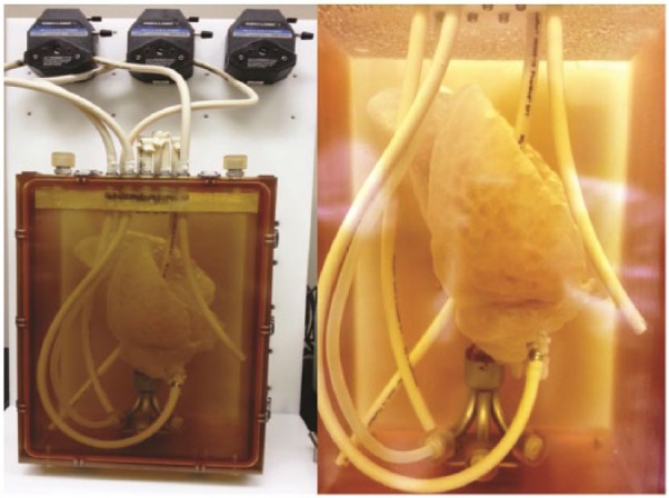
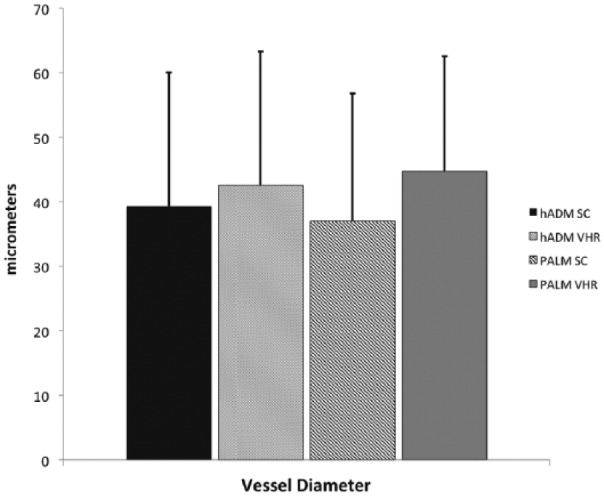
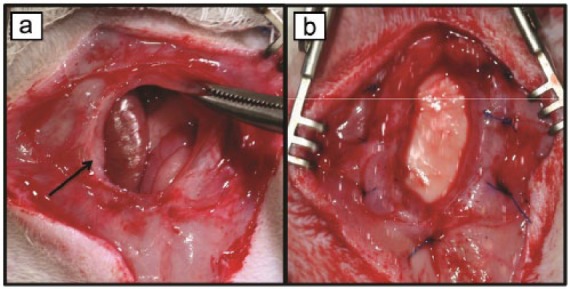
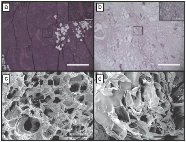
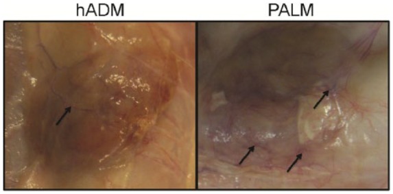
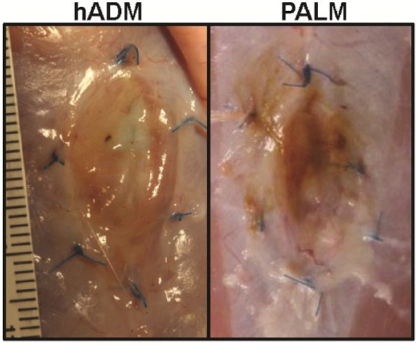
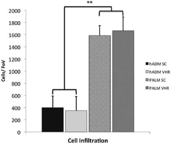
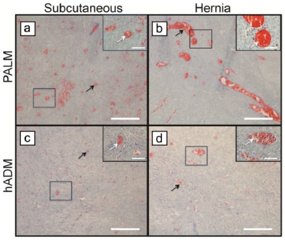
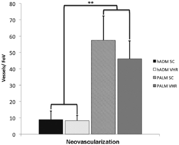
Surgical wound healing applications require bioprosthetics that promote cellular infiltration and vessel formation, metrics associated with increased mechanical strength and resistance to infection. Porcine acellular lung matrix is a novel tissue scaffold known to promote cell adherence while minimizing inflammatory reactions. In this study, we evaluate the capacity of porcine acellular lung matrix to sustain cellularization and neovascularization in a rat model of subcutaneous implantation and chronic hernia repair. We hypothesize that, compared to human acellular dermal matrix, porcine acellular lung matrix would promote greater cell infiltration and vessel formation. Following pneumonectomy, porcine lungs were processed and characterized histologically and by scanning electron microscopy to demonstrate efficacy of the decellularization. Using a rat model of subcutaneou implantation, porcine acellular lung matrices (n = 8) and human acellular dermal matrices (n = 8) were incubated in vivo for 6 weeks. To evaluate performance under mechanically stressed conditions, porcine acellular lung matrices (n = 7) and human acellular dermal matrices (n = 7) were implanted in a rat model of chronic ventral incisional hernia repair for 6 weeks. After 6 weeks, tissues were evaluated using hematoxylin and eosin and Masson's trichrome staining to quantify cell infiltration and vessel formation. Porcine acellular lung matrices were shown to be successfully decellularized. Following subcutaneous implantation, macroscopic vessel formation was evident. Porcine acellular lung matrices demonstrated sufficient incorporation and showed no evidence of mechanical failure after ventral hernia repair. Porcine acellular lung matrices demonstrated significantly greater cellular density and vessel formation when compared to human acellular dermal matrix. Vessel sizes were similar across all groups. Cell infiltration and vessel formation are well-characterized metrics of incorporation associated with improved surgical outcomes. Porcine acellular lung matrices are a novel class of acellular tissue scaffold. The increased cell and vessel density may promote long-term improved incorporation and mechanical properties. These findings may be due to the native lung scaffold architecture guiding cell migration and vessel formation. Porcine acellular lung matrices represent a new alternative for surgical wound healing applications where increased cell density and vessel formation are sought.
外科伤口愈合应用需要能够促进细胞浸润和血管形成的生物假体,这些指标与机械强度增加和抗感染能力相关。猪脱细胞肺基质是一种新型组织支架,已知其能促进细胞黏附,同时将炎症反应降至最低。在本研究中,我们评估了猪脱细胞肺基质在大鼠皮下植入和慢性疝修补模型中维持细胞化和新血管形成的能力。我们假设,与人类脱细胞真皮基质相比,猪脱细胞肺基质能促进更大程度的细胞浸润和血管形成。肺切除术后,对猪肺进行处理,并通过组织学和扫描电子显微镜进行表征,以证明脱细胞效果。使用大鼠皮下植入模型,将猪脱细胞肺基质(n = 8)和人类脱细胞真皮基质(n = 8)在体内孵育6周。为了评估在机械应力条件下的性能,将猪脱细胞肺基质(n = 7)和人类脱细胞真皮基质(n = 7)植入大鼠慢性腹直肌切口疝修补模型中6周。6周后,使用苏木精和伊红以及Masson三色染色对组织进行评估,以量化细胞浸润和血管形成。结果表明猪脱细胞肺基质成功脱细胞。皮下植入后,宏观血管形成明显。猪脱细胞肺基质显示出充分的整合,并且在腹疝修补后没有机械故障迹象。与人类脱细胞真皮基质相比,猪脱细胞肺基质显示出明显更高的细胞密度和血管形成。所有组的血管大小相似。细胞浸润和血管形成是与改善手术结果相关的整合的良好表征指标。猪脱细胞肺基质是一类新型的脱细胞组织支架。细胞和血管密度的增加可能促进长期改善的整合和机械性能。这些发现可能是由于天然肺支架结构引导细胞迁移和血管形成。猪脱细胞肺基质代表了一种新的外科伤口愈合应用替代方案,适用于需要增加细胞密度和血管形成的情况。